Tales From Honduras – Ichthyosis, An Unusual Skin Finding
Case
While working in a rural mountain village in Honduras, a 4-year-old boy presents to clinic accompanied by his grandmother. He has no acute complaints apart from decreased hearing in his left ear for the past week. There has been no pain, discharge, fevers, or other associated symptoms. Additionally, his grandmother makes note of a skin “rash” he has had since birth. Otherwise, he has been growing well, eating normally, and occasionally suffers from dry eyes. He has a family member with a similar, but less dramatic skin finding.
On examination, he has normal vital signs. He is in no distress, comfortable appearing, but acting shy and withdrawn (appropriate for age). His eyes and conjunctiva are normal, but the eyelids are ectropic. He has moist and otherwise normal oral mucosa. The left otic canal is filled with keratinitic debris and once cleared reveals a normal tympanic membrane. The right ear is normal. The remainder of his skin examination reveals diffuse, whole-body brown plaques which are anchored at the center and peeling at the edges, no erythema, purulent drainage, or tenderness to palpation. He has alopecia. The remainder of his examination was normal.


Diagnosis
Ichthyosis, most likely autosomal recessive congenital ichthyosis (ARCI), lamellar type.
Discussion
Generally speaking, ichthyoses are a set of predominantly genetic disorders affecting skin barrier formation resulting in the characteristic dry skin, scaling, and hyperkeratosis. These disorders are caused by a myriad of genetic defects and are broadly characterized into syndromic and non-syndromic varieties, with the latter being confined primarily to skin manifestations. The most common forms of non-syndromic forms are ichthyosis and recessive X-linked ichthyosis while the less common variants include autosomal recessive congenital ichthyosis and keratinopathic ichthyosis.[1,2]
Ichthyosis vulgaris (incidence 1:250 – 1:1000)
This is the mildest form and is characterized by mild scaling, pruritis, eczema and xerosis. There is often seasonal variation, with improvement in the warmer months. Manifestations are usually limited to the extremities.[1]
Ichthyosis Vulgaris. Courtesy of Contour Dermatology
Recessive X-linked Ichthyosis (incidence 1:2000 – 1:6000)
This is the second most common ichthyosis and is often present from birth. It can be distinguished by generalized xerosis, large, rhomboid, dark brown scales across the entire body, and no seasonal variation.[1]

Recessive X-linked Ichthyosis courtesy of DermNet NZ.
Autosomal Recessive Congenital Ichthyosis (incidence 1:200,000)
This entity exists on a continuum which includes congenital ichtyosiform erythroderma (CIE), lamellar, and harlequin type, as well as multiple minor subtypes.[1-4]
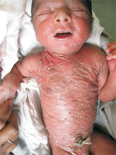
CIE is the least severe form of ARCI and patients have the most intact skin barrier throughout life. At birth, CIE patients are covered by a collodion membrane (see bottom for more information). They later progress to develop a fine white scaling overlying a persistent, generalized erythroderma.[1,2,4]
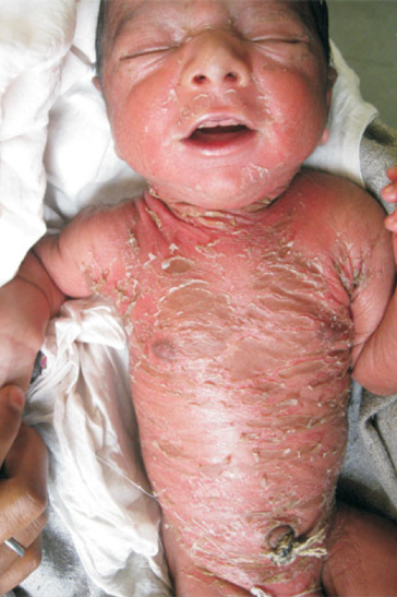
Infant with remnants of a collodion membrane and generalized erythroderma suggesting the diagnosis of CIE

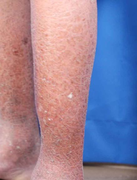
Adult patient with CIE
The lamellar type of ARCI is a less severe form than the harlequin type, but it puts patients at increased susceptibility to infection (particularly as an infant) due to disruption of the skin barrier. These patients will have a collodion membrane at birth and then progress to develop large, brown plate like scales with large fissures between them. Alopecia as well as palmar/plantar hyperkeratosis are commonly seen. Because of the large fissures lamellar type ARCI patients are at high risk of superinfection, as well as heat related illness due to hypohydrosis from disrupted sweat glands.[1,2,4]

Figure 10. Representative drawing of Lamellar Ichthyosis showing the typical “dry riverbed” appearance of the skin.
Harlequin ichthyosis is the most severe and by far the most dramatic form of ARCI. Harlequin ichthyosis carries up to a 50% neonatal mortality rate. This high mortality is attributed to overwhelming infections from skin barrier effects, respiratory failure from skin tightness (similar to the restrictive pathology of burn eschars), and malnutrition from an inability to take oral nutrition. The disease process is characterized by premature birth, ectropion (eyelid eversion), eclabium (lip eversion), auricular malformation, and a characteristic “armor plate” keratosis. Unfortunately, treatment is largely supportive and best administered in a neonatal intensive care unit although there is growing evidence for the use of retinoids in the neonatal period.[1,2,4]
![Figure 11. A neonate with the typical appearance of harlequin ichthyosis (left) which, in adulthood, (right) can later resemble congenital ichthyosiform erythroderma.[ Kün-Darbois, then daily mail]](https://images.squarespace-cdn.com/content/v1/56e8a86a746fb97ea9d14740/1587647559607-YEES2SXWQCS2GJDUYFV9/Screen+Shot+2020-04-23+at+8.12.18+AM.png)
Figure 11. A neonate with the typical appearance of harlequin ichthyosis (left) which, in adulthood, (right) can later resemble congenital ichthyosiform erythroderma.[ Kün-Darbois, then daily mail]
Treatment and Diagnosis Considerations
Diagnosis is typically clinical and based on history, timing, and exam findings. While biopsy and genetic testing are available, they are not always necessary or available.[1-4]
Type and intensity of therapy should be titrated to disease severity. For more mild cases, skin hydration can be achieved with topical creams and ointments. For more severe cases, systemic treatment with oral retinoids should be considered for their keratinolytic effects. However, these are high risk medications with teratogenic effects and appropriate precautions must be taken. Lifestyle modifications include daily bathing with mild cleansers, frequent application of topical emollients, and avoidance of environmental factors, both of which can be financially and temporally burdensome to patients. Finally, genetic counseling is advised for those patients considering children.[2,4,5]
Case Resolution
Ultimately, the patient was discharged with moisturizing lotion, artificial tears, and Vaseline for the face. He was encouraged to follow closely with the nearby health clinic for skin checks by the nurse. Lastly, he was counseled to maintain good nutrition, hydration, and to avoid excessive heat.
Take-Aways
There are many different forms of ichthyosis, which have a wide range of disease severity.
Complications can arise from breakdown of the skin barrier resulting in super-infection, fluid losses, and heat related illness.
Treatment focuses on skin hydration and lifestyle modifications. While oral agents can be used, they are best administered under the guidance of specialists because of associated risks.
Medical Trivia -- Collodion Membrane
Derived from the Greek word for glue, a collodion membrane refers to a thin sac of adherent keratinocytes that enclose some infants. This thin, clear, “cellophane-like” membrane can persist for two days up to two weeks. Infants appear to be glistening as if they were “dipped in wax.” The presence of this membrane indicates a high risk of developing ichthyosis (~50%) and a risk of developing one of the syndromic keratosis.[2,6]

Infant with the typical “cellophane wrapped” appearance of a collodion membrane.
Faculty reviewer: Kyle Martin, DO, MA, MPH, DTM&H
References
Takeichi T, AkiyamaM. Inherited ichthyosis: Non‐syndromic forms. J Dermatol. 2016; 43:242-251.
Vahlquist A, Fischer J, Törmä H. Inherited Nonsyndromic Ichthyoses: An Update on Pathophysiology, Diagnosis and Treatment. Am J Clin Dermatol. 2018;19(1):51–66.
Richard G. Autosomal Recessive Congenital Ichthyosis. 2001 Jan 10 [Updated 2017 May 18]. In: Adam MP, Ardinger HH, Pagon RA, et al., editors. GeneReviews® [Internet]. Seattle (WA): University of Washington, Seattle; 1993-2018. Available from: https://www.ncbi.nlm.nih.gov/books/NBK1420/
Craiglow BG. Ichthyosis in the newborn. Semin Perinatol. 2013;37(1):26–31.
Limmer A, Nwannunu C, Patel R, Mui U, Tyring S. Management of Ichthyosis: A Brief Review. Skin Therpay Lett. 2020; 25(1):5-7.
Phuljhele S, Hura KS, Khandwal O. Collodion baby: A case report. Inter J Medical Sci Res Prac 2015; 2(2):102-103.
Images
(ichtyosis vulgaris) Contour Dermatology [Internet]. Ichthyosis vulgaris. 2018 [accessed 2020 April 16]. Available from: https://contourderm.com/ichthyosis-vulgaris/
(Recessive X-linked Ichthyosis) DermNet NZ [Internet]. Recessive X-linked Ichthyosis. 2015 [accessed 2020 April 16]. Available from: https://dermnetnz.org/topics/recessive-x-linked-ichthyosis/
(harlequin infant) Kün-Darbois JD, Molin A, Jeanne-Pasquier C, Paré A, Bénateau H, Veyssière A. Facial features in Harlequin ichthyosis: Clinical findings about 4 cases. Revue de Stomatologie, de Chirurgie Maxillo-faciale et de Chirurgie Orale. 2016; 117(1):51-53.
(Harlequin adult) Daily Mail [Internet]. 'Snakeskin woman', 33, becomes the oldest ever with a rare condition that causes her skin to grow thick and scaly (and she wants you to know she's just like anyone else). 2017 [accessed 2020 April 16]. Available from: https://www.dailymail.co.uk/femail/article-4337800/Snakeskin-woman-oldest-rare-skin-condition.html
(lamellar diagram) List of Disease [Internet]. Lamellar Ichthyosis. 2011 [accessed 2020 April 16]. Available from: https://listofdisease.wordpress.com/2011/10/03/lamellar-ichthyosis/
(collodion baby) Medical Pictures Info [Internet]. Collodion Baby. 2011 [accessed 2020 April 16]. Available from: https://medicalpicturesinfo.com/collodion-baby/
(CIE baby) Indian Pediatrics [Internet]. 2014 [accessed 2020 April 16]. Available from:
(adult CIE) Danderm [Internet]. Atlas of Clinical dermatology. 2015 [accessed 2020 April 16]. Available from: http://www.danderm-pdv.is.kkh.dk/atlas/pics/5/5-61-3.jpg