Article

Fragaria × ananassa cv. Senga Sengana Leaf: An Agricultural Waste with Antiglycation Potential and High Content of Ellagitannins, Flavonols, and 2-Pyrone-4,6-dicarboxylic Acid

Department of Pharmacognosy and Herbal Medicines, Faculty of Pharmacy, Wroclaw Medical University, Borowska 211a, 50-556 Wroclaw, Poland
*
Author to whom correspondence should be addressed.
Molecules 2022, 27(16), 5293; https://doi.org/10.3390/molecules27165293
Submission received: 31 July 2022 / Revised: 16 August 2022 / Accepted: 16 August 2022 / Published: 19 August 2022
(This article belongs to the Special Issue Phytochemistry and Biological Properties of Medicinal Plants)

Abstract

:
Strawberry leaves are considered a valuable waste material; so far, mainly due to their antioxidant properties. Since the annual production of this crop is high, our study aimed to thoroughly examine the chemical composition and antidiabetes-related bioactivity of Fragaria × ananassa leaf of its popular and productive cultivar Senga Sengana. Leaves from three different seasons, collected after fruiting, were extensively analyzed (UHPLC-qTOF-MS/MS, HPLC-DAD). Some individual components were isolated and quantified, including specific flavonol diglycosides (e.g., 3-O-[β-xylosyl(1‴→2″)]-β-glucuronosides). The separated quercetin glycosides were tested in an antiglycation assay, and their methylglyoxal uptake capacity was measured. In addition, the biodegradable polyester precursor 2-pyrone-4,6-dicarboxylic acid (PDC) was confirmed at relatively high levels, providing further opportunity for strawberry leaf utilization. We want to bring to the attention of the food, pharmaceutical, and cosmetic industries the Senga Sengana strawberry leaf as a new botanical raw material. It is rich in PDC, ellagitannins, and flavonols—potent glycation inhibitors.

Graphical Abstract

1. Introduction

Plant polyphenols provided to the human body with herbs, vegetables, or fruits significantly influence the digestive system, internal organs, and tissue activity. However, their bio-accessibility is diverse and conditioned by their chemical structure. The available epidemiological and interventional studies featuring healthy volunteers and patients show that polyphenols with antioxidative, antiglycative, and antiphlogistic properties reduce the risk of some chronic noncommunicable diseases, such as diabetes, metabolic syndrome, fatty liver disease, cardiovascular disease, and some cancers. Undoubtedly, in the etiology of those illnesses, an important role is played by pathogenic oxidative and carbonyl stress and ongoing inflammation [1,2,3]. In this regard, much attention has been paid to the properties of commonly consumed polyphenols [4,5]. There is also ongoing research on the interactions between polyphenols and gut microbiota to elucidate their health benefits. The data on flavonoids and ellagitannins are of particular interest [6,7].
In traditional medicine, leaves of species providing berry fruits (such as blackberry, raspberry, or wild strawberry) were used to cure gastroenteritis and mild diarrhea, strengthen the heart and circulatory system, and in metabolic disturbances to ‘improve metabolism’ and for ‘blood purification.’ The effectiveness of those recommendations is not sufficiently documented; therefore, their therapeutic application is currently limited. For example, the wild strawberry leaf is included in several multi-component food supplements and traditional herbal medicines (traditional botanical drugs) in the EU, intended to relieve non-specific diarrhea or enhance diuresis and metabolism. Similarly, it is used as a component of notified and licensed products in the USA and Canada [8]. Herbal medicines containing wild strawberry leaf are usually available as herbal teas for oral use.
It should be noted that wild strawberry leaf is the common name shared by the dried leaves of at least four species: Fragaria vesca L., F. moschata Weston, F. viridis Weston, and F. ananassa (Weston) Duch. ex Rozier. This raw plant material is described, inter alia, in the European Union herbal monograph Fragariae folium (EMA/HMPC/432278/2015). Leaves of Fragaria species with or without petioles can be harvested throughout the growing season. However, according to older data, wild strawberry leaf for therapeutic purposes was collected during the flowering period [8].
Phytochemical studies have shown a similar chemical profile of leaves from Fragaria species [9]. Nevertheless, scientific references focus mainly on F. vesca (woodland strawberry). The woodland strawberry leaf contains up to 12% tannins (ellagitannins and proanthocyanidins), 0.2–4% flavonoids (glycosides of flavonols), phenolic acids (ellagic acid, gallic acid, hydroxycinnamic acids), triterpenes, traces of essential oil, and others [10]. Its main components are quercetin, kaempferol glycosides, catechin, and agrimoniin [8,11]. The chemical identity of many others has not been revealed yet.
Due to the efforts of growers and the adaptability of F. ananassa plants, strawberry cultivation has spread almost worldwide. According to FAO data, world strawberry production in 2020 was nearly 7 million tonnes, and the top three global strawberry producers were China, the USA, and Egypt [12]. The Senga Sengana strawberry has been a leading industrial cultivar for many years. It is characterized by abundant foliage, consisting of large, shiny leaves with a dark green surface. The older leaves are removed in the crops about 2–3 weeks after harvesting the berries by pruning all the plants (July/August). The treatment is applied to 2- and 3-year-old strawberries. The plant material remaining after this procedure has unexplored potential for utility.
For these reasons, in the present study we performed a detailed examination of the chemical composition of F. ananassa cv. Senga Sengana leaves gathered after fruiting as regards polyphenols (including tannins, flavonoids, phenolic acids) and carboxylic acids. The results obtained for Senga Sengana leaves were compared to leaves of F. vesca, a research model used in other studies. Since flavonoids are characterized by their ability to take up methylglyoxal and inhibit the destructive glycation of bio-molecules [13], we decided to compare the effects of isolated quercetin glycosides with known glycation inhibitors such as aminoguanidine and metformin. Based on this, we identified a new botanical raw material with valuable phytochemicals and high utility potential.

2. Results

The leaves of species from the Fragaria genus are considered rich in antioxidants and thus of interest to the food, pharmaceutical, and cosmetic industries. Among them, garden strawberry leaves are the most broadly available renewable resource that can be harvested yearly after fruiting. There are many cultivars of garden strawberry; however, we do not know the complete chemical composition of most of them—neither the fruit nor the leaf.
The Senga Sengana strawberry has been consistently touted as a leading industrial cultivar for many years. It was first bred in Germany, and today it performs widely in commercial and amateur cultivation.
The examined leaves of F. ananassa cv. Senga Sengana were collected in July of three different years from a local plantation (FaSS1, FaSS2) and the experimental farm COBORU (FaSS3). Preliminary chromatographic tests of 50% water-methanol extracts of them revealed a significant content of polar polyphenolic metabolites. Among other components, the presence of numerous flavonoids and hydrolyzable tannins was confirmed. However, we were unable to confirm the identity of several peaks with certainty, and this became one of the targets of the present experiment. Therefore, the dried leaves of FaSS1 were intended to isolate tannins, flavonoids, and derivatives of phenolic and carboxylic acids, whereas FaSS3 leaves were used for final UHPLC-qTOF-MS/MS profiling (Table 1). Since the diploid woodland strawberry (F. vesca) is frequently used to study the more complex octoploid garden strawberry (F. ananassa), we compared the leaf chemistry of the Senga Sengana strawberry with two woodland strawberry reference samples (Fv1, Fv2).
In-depth investigation of F. ananassa cv. Senga Sengana leaf polyphenols was carried out using various chromatographic (CC, TLC, HPLC-DAD, UHPLC-qTOF-MS/MS) and spectroscopic methods: ultraviolet to visible light spectroscopy (UV-VIS), electrospray ionization mass spectrometry (ESI-qTOF-MS), as well as one-dimensional (1H-, 13C-NMR, DEPT) and two-dimensional nuclear magnetic resonance spectroscopy (COSY, NOESY, HSQC, HMBC). In addition, plant extracts subjected to qualitative and quantitative analysis were prepared from leaves of both garden and woodland strawberries, using a procedure described previously [11].
A vital step to characterize the polyphenols of F. ananassa cv. Senga Sengana leaf was the separation of individual compounds on a preparative scale. Dried leaves of FaSS1 (500 g) were exhaustively extracted with water-acetone (1+1). The obtained aq. acetone extract (FaSS-A) was concentrated and fractionated using column chromatography on octadecyl, silica gel Si60 and Sephadex LH-20. From the octadecyl column, we obtained 13 fractions rich in polyphenols and other components (FaSS-0, FaSS-I to FaSS-XII), which were purified on silica gel Si60 (FaSS-I) or Sephadex LH-20 (FaSS-0, FaSS-II to FaSS-IX). FaSS-X and FaSS-XI, containing one dominant component each, were concentrated and allowed to crystallize from water-methanol (Figure S1). All isolated compounds were identified using authentic standards and tannins previously separated by the authors from various sources (if applicable; co-chromatography: TLC on silica gel 60 in mobile phases E, F, P—comparison of Rf, color, and its intensity; HPLC-DAD—tR, UV-VIS spectra—λmax and absorption profile) (Figures S2–S5), and characterized by ESI-qTOF-MS/MS in negative mode. In addition, 1D- and 2D-NMR experiments were used for structural elucidation of hydrolyzable tannins, flavonoid glycosides, and derivatives of phenolic and carboxylic acids separated from FaSS-A (see Appendix A and Appendix B for isolation/identification details and Supplementary Materials for MS/MS and NMR details).
To summarize, sixteen individual compounds were isolated from the aq. acetone extract of Senga Sengana leaves. These included four hydrolyzable tannins, compounds 3 (3-O-galloylquinic acid) [14,15], 5 (5-O-galloylquinic acid) [14,15], 7 (pedunculagin) [16], and 10 (agrimoniin) [17,18]; seven flavonoid glycosides, compounds 8 ((2R,3R)-taxifolin-3-O-β-glucoside) [19], 9 (quercetin-3-O-β-glucuronoside-7-O-β-glucoside) [20], 11 (quercetin-3-O-[β-xylosyl(1‴→2″)]-β-glucuronoside) [21], 12 (quercetin-3-O-β-glucuronoside) [22], 13 (kaempferol-3-O-[β-xylosyl(1‴→2″)]-β-glucuronoside, newly described), 14 (isorhamnetin-3-O-β-glucuronoside) [23], and 15 (tiliroside) [24]; and five other compounds: 1 (2-pyrone-4,6-dicarboxylic acid, PDC) [25], 2 (quinic acid), 4 (4a: 1-O-protocatechuoyl-β-xylose, newly described), 6 (catechin) [26], and 16 (ellagic acid).
To identify minor Senga Sengana components, we performed UHPLC-qTOF-MS/MS profiling with co-chromatography (Table 1). Figure 1 shows a typical HPLC-DAD chromatogram of 50% water-methanol extract from leaves of F. ananassa cv. Senga Sengana. The structures of main ellagitannins identified in the analyzed leaves are presented in Figure 2, those of flavonoids in Figure 3, while other are depicted in Figure 4.

2.1. Hydrolyzable Tannins

Agrimoniin (10) was previously obtained from Agrimonia pilosa Ledeb. (syn. A. japonica (Miq.) Koidz.) and Potentilla kleiniana Wight et Arnott. [17,18,29], as well as from F. ananassa Duch. and F. nipponica Makino leaves [29], and berries of woodland and garden strawberry [45]. The content of agrimoniin in plant materials FaSS1 and FaSS2 was determined as ca. 4.4% and 3% (dry matter), respectively, while it was 8.1% in FaSS3 (Table 2). Pedunculagin (7) was previously reported in strawberry leaves by Okuda et al. [29].
Monogalloylquinic acids 3 and 5 (classified in gallotannins) were identified as 3- and 5-O-galloylquinic acid, respectively [14,15]. The content of dominant 5-O-galloylquinic acid in Senga Sengana leaves ranged from 0.4 to 0.6% of dry matter (Table 2). The other regioisomers of monogalloylquinic acid (Table 1) were present in much lower amounts.
The identity of other minor ellagitannins (ETs: davuriciin D2/fragariin A; laevigatins B/C/F; potentillin; galloyl-HHDP-glucose isomers such as sanguiin H-4; digalloyl-HHDP-glucose), as well as gallotannins (3,5-O-digalloylquinic acid; mono-, di- and trigalloylglucose) [27,32,34,35,36,37] in strawberry leaf extracts and fractions was determined using UHPLC-qTOF-MS/MS (Table 1). Davuriciin D2 was isolated for the first time from the root of Rosa davurica Pall. [37]. Ellagitannin with MW 2038 Da was also obtained from strawberry fruit pomace under the name fragariin A [32]. Analysis of the published spectroscopic data and structures proposed for these ETs by their authors indicates that they are most likely identical. In addition, four ETs with molecular weights of 934 (two isomers), 1236, and 2020 Da were detected in Senga Sengana leaves. The same compounds were observed in pseudo-fruits, flowers, and leaves of several other F. ananassa cultivars, but their formulas were not elucidated [27,28,30,36]. Fragmentation of those minor strawberry ETs in the negative ion mode provided products typical for HHDP esters (m/z 481 and 301) and an additional ion derived from a low molecular depside at m/z 451 (exactly 450.9950). The same signal has been observed on the MS2 spectrum of davuriciin D2/fragariin A (MW 2038 Da, m/z 1018.0749 [M−2H]−2), which is a valoneoyl analog of agrimoniin [37]. Therefore, the presence of the m/z 451 ion suggests a structural affinity, so we tentatively propose their chemical formulas starting with davuriciin D2/fragariin A (dimeric ellagitannin composed of α-pedunculagin and α-praecoxin A linked by DHDG, Figure 2). Ellagitannin-2020 is possibly a lactone of davuriciin D2/fragariin A (in the valoneoyl group) composed of α-pedunculagin and α-praecoxin D coupled by DHDG. The remaining compounds are secondary ellagitannins formed by the neutral loss of monomeric ellagitannin molecules, i.e., davuriciin D2/fragariin A lactone without the pedunculagin fragment (ET-1236 → 2020 − 784 = 1236) and davuriciin D2/fragariin A lactone without pedunculagin and DHDG coupler (ET-934 isomers → 2020 − 784 − 302 = 934). The latter compounds are probably α- and β-anomers of praecoxin D [31]. The absence of the DHDG lactone-derived ion in MS2 supports that thesis. As a result of the fragmentation of m-GOG type dimers and certain secondary ellagitannins, besides both ellagic acid (m/z 301) and decarboxylated monolactone of hexahydroxydiphenic acid (LHHDP−CO2, m/z 275) ions, a lactone of the dehydrodigallic acid ion (LDHDG, m/z 319) also occurred. Tannins with a valoneoyl group additionally released the diagnostically relevant ion of valoneic acid trilactone (VTL, m/z 451). The reported ETs with molecular weights below 1870 Da were probably products of partial degradation or metabolism of agrimoniin and davuriciin D2/fragariin A. D2 content was many times lower than agrimoniin at 0.2–0.8% of dry matter (Table 2).
The quantitative analysis of ellagitannins in various parts of strawberry cultivars in relation to the development stage was the topic of work of Karlińska and co-workers [46].

2.2. Proanthocyanidins and Flavan-3-Ols
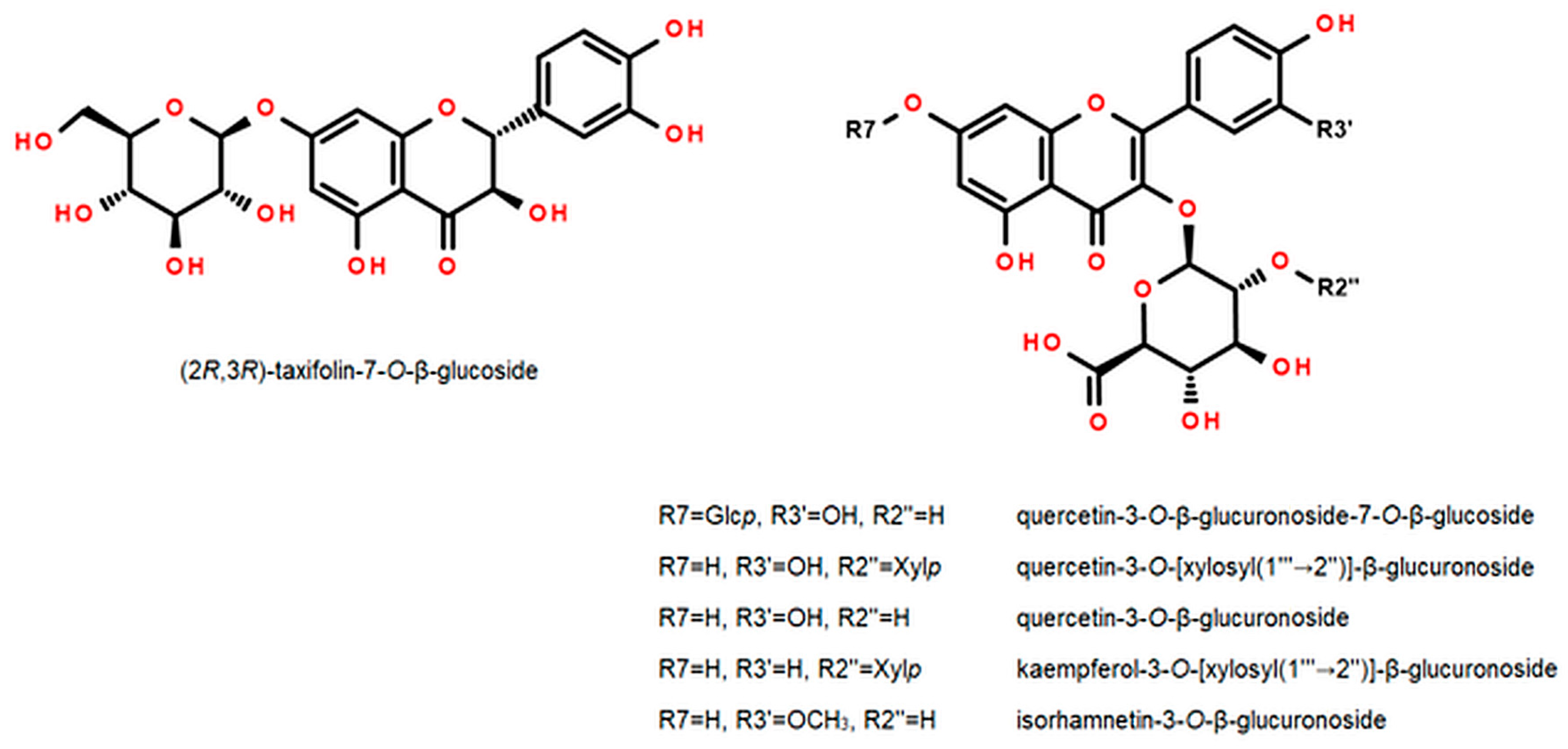
Condensed tannins were minor F. ananassa cv. Senga Sengana components. Several B-type proanthocyanidins (five dimers, three trimers, and one tetramer) were detected together with monomeric catechin by UHPLC-qTOF-MS/MS (m/z 289.0719, 577.1346, 865.1981, 1153.2610). Except for catechin (6), procyanidins B3 and C2, as well as oligomeric flavan-3-ol derivatives of both catechin and epicatechin, were recognized by UV-VIS profiles and MS fragmentations. Catechin and procyanidins B3 and B6 were formerly isolated from F. ananassa cv. Reikov roots [26].

2.3. Flavonoids

Seventeen flavonoid glycosides were recognized in extracts from Senga Sengana leaves (Table 1, Figure 3). Five compounds were derivatives of quercetin, five of kaempferol, one of isorhamnetin, and six were taxifolin glycosides. In this study, we separated the dominant flavonoids, especially those that have not been further characterized. Three diglycosides (9, 11, and 13), as well as four monoglycosides (8, 12, 14, and 15), were isolated from FaSS-A. Some of the noted flavonoids, including flavonol diglycosides (MW 594, 610, and 640 Da), were described previously in leaves of F. ananassa cv. Polka [27], in flowers of the cultivar Jonsok [28], and in pseudo-fruits of the Japanese cultivar Tochiotome [21]. Nevertheless, the abovementioned authors assigned the structures only tentatively by LC-MS.
The NMR data of flagarin (11) were in accordance with those published data [21]. The mentioned authors reported the presence of 3-O-β-(2″-Xylp)-GlcAp of quercetin in strawberry fruits. However, the final semi-systematic name of this compound was given by them inversely in the sugar part, despite clear proof. Our results indicate that compound 11 should be quercetin-3-O-[β-xylosyl(1‴→2″)]-β-glucuronoside.
Compound 13 (yellow crystalline powder) had a pseudomolecular ion at m/z 593.1144 [M−H] (calc. 593.1148 for [C26H25O16]), as well as a fragment ion at 285 [M−308−H] (aglycone). These data were relevant to hexuronoside-pentoside or rutinoside of kaempferol-like aglycone. Flavonoid 13 showed an analogic fragmentation pattern to 11, so we deduced that sugars should be attached sequentially. After acid hydrolysis, 13 yielded glucuronic acid, xylose (mobile phase S), and kaempferol. 13C-NMR (DMSO-d6) data showed signals strictly corresponding to a glucuronosyl and a xylosyl, similarly as in 11, but kaempferol was proved as an aglycone (doubled singlets of H-2′ & H-6′, and H-3′ & H-5′ in ring B). 1H-NMR showed precisely two doublets of anomeric protons (4.61 and 5.73 ppm), of which the shift with higher value was assigned to β-GlcAp. The glycosylation position was deduced similarly as in 11, from C-3 upfield shift from ~135 to ~132 ppm together with downfield C-4 and C-1′ signals. The second sugar of glycone (β-Xylp) was attached to the first one in the same way as in 11. That was proved by 2D-NMR (NOESY: H-1‴↔H-2″) and downfield of the C-2″ signal of the β-GlcAp moiety (from ~73 to ~81 ppm). The NMR data were closely similar to 11, an analogic glycoside of quercetin [21]. Our results indicate that compound 13 should be 3-O-[β-xylosyl(1‴→2″)]-β-glucuronoside of kaempferol. This compound is newly described and supplied with NMR data (Appendix B, Supplementary Materials).
All spectroscopic data of 8 were clearly similar to those included in a previous paper [19] and different from those of Pan and Lundgren [47], but the spectrum revealed several overlapping isomer signals. That compound was also found in Tochiotome strawberries [21]. Other taxifolin pentosides and hexosides were also observed in LC-MS as minor components (Table 1). For example, taxifolin-3-O-α-arabinoside was isolated previously from roots of F. ananassa cv. Reikov [26].
Based on spectroscopic evidence, flavonoids 12, 14, and 15 were identified as quercetin-3-O-β-glucuronoside (syn. Quercetin-3-O-β-glucuronide or miquelianin, m/z 477.0672), isorhamnetin-3-O-β-glucuronoside (3′-O-methyl-quercetin-3-O-β-glucuronoside, 491.0806), and tiliroside (kaempferol-3-O-β-(6″-O-p-coumaroyl) glucoside). Kaempferol, quercetin, and isorhamnetin-3-O-(6″-methyl) glucuronosides were formerly identified in fruits of F. ananassa cv. Falandi [48], whereas cis/trans tiliroside was identified in leaves of cv. Jonsok [42] and in pseudo-fruits of cultivars Tochiotome [41], Nohime [39], and Minomusume [40]. The structures of the most important flavonoids are presented in Figure 3.
The most abundant compound of the flavonoid fraction was quercetin-3-O-[β-xylosyl(1‴→2″)]-β-glucuronoside (11, flagarin ∼0.7%), followed by kaempferol-3-O-[β-xylosyl(1‴→2″)]-β-glucuronoside (13, ∼0.5%), quercetin-3-O-β-glucuronoside (12, 0.1–0.3%) and quercetin-3-O-β-glucuronoside-7-O-β-glucoside (9, ∼0.05%) (Table 2). The sum of flavonol glycosides in Senga Sengana leaves was determined at 1.5–1.7% (dry matter). Kårlund and coworkers quantified the quercetin and kaempferol glycosides in leaves of the strawberry cultivar Polka [27] at concentrations comparable to our study.

2.4. Phenolic and Carboxylic Acids

UHPLC-qTOF-MS/MS analysis of F. ananassa cv. Senga Sengana leaf extracts showed intense peaks of several phenolic acid derivatives, as well as carboxylic acids. Among them, 1 was identified as 2-pyrone-4,6-dicarboxylic acid (PDC, Figure 4). This heterocyclic dicarboxylic acid is considered as a chemotaxonomic marker of Rosoideae and was formerly reported in a concentration of 0.1–2% in the following genera: Alchemilla, Agrimonia, Duchesnea, Filipendula, Fragaria, Geum, Potentilla, Rosa, Rubus, Sanguisorba, and Waldsteinia [25]. PDC is also known as a low molecular product of catabolism (biodegradation) of plant polyphenols (phenolic acids, lignans, and lignin) by some microorganisms, e.g., by the soil bacterium Sphingomonas paucimobilis [49]. The biological origin of 2-pyran-4,6-dicarboxylic acid in strawberry leaves has not been definitively elucidated. In the examined raw plant material, the PDC content was in the range of 1–1.7% dry matter (Table 2). Carboxylic acid 2, obtained from the same fraction as PDC, was identified as a cyclic polyol named quinic acid. The presence of citric acid was also confirmed in Senga Sengana leaves (Table 1).
Compound 4a was separated as an off-white crystalline powder from fraction FaSS-I. Its pseudomolecular ion m/z at 285.0614 [M−H] (calc. 285.0616 for [C12H13O8]) was relevant to O-pentoside of dihydroxybenzoic acid or dihydroxybenzoyl-pentose. Compound 4a produced both radical aglycone (Y0−H) and aglycone Y0 ions at m/z 152 and 153 (by homolytic and heterolytic cleavage of the deprotonated precursor ion), as well as at 108 and 109 [50], corresponding to a dihydroxybenzoic acid residue after the loss of pentose followed by decarboxylation. The ions formed by the homolytic cleavage were dominant, which suggested the presence of two free phenolic groups in the benzene ring. In the 1H-NMR spectrum of 4, a dominant anomeric signal (4a) was accompanied by an additional small one (4b). The 13C-NMR spectrum of 4a showed signals corresponding to β-Xylp of protocatechuic acid. Shifts of glycone carbons were compared positively with literature data [51]. Esterification with protocatechuic acid was confirmed by simple acidic and alkaline hydrolysis, as Markham described [51]. Our results indicate that 4a should be 1-O-protocatechuoyl-β-xylose (Figure 4). As an impurity of 4a, protocatechuic acid-3-O-β-glucoside (4b) was deduced.
Besides EA (16), O-glycosides of ellagic acid were distinguished in Senga Sengana leaf extracts (m/z 433.0408 and 447.0570). Ellagic acid glycosides fragmented to ions corresponding to EA at m/z 301 and its decarboxylated monolactone at 275 [LHHDP−44/CO2−H]. Analogous pentosides and deoxyhexosides of ellagic acid and methylellagic acid were identified in strawberries from cultivars Alba, Clery, Darselect, Elsanta, Eva, Portola [44], and others.
The next group of compounds characterized by UHPLC comprised phenylethanoids (isomers of 1-O-β-[2′-(4″-hydroxyphenyl)ethyl]-6-O-(p-coumaroyl)glucose) with a pseudomolecular ion [M−H] at m/z 445.1507 (calc. 445.1504 for [C23H25O9]) and fragment ions characteristic for p-coumaric acid esters: 163 [p-coumaric acid−H], 145 [p-coumaroyl−H] and 119 [p-coumaric acid−44/CO2−H]. Their UV-VIS spectra exhibited λmax at 312 nm, resulting from the HCA moiety. A compound with a pseudomolecular ion 461.1448 (calc. 461.1453 for [C23H25O10]) and λmax at 323 nm was tentatively identified as a caffeoyl ester of hydroxyphenylethanol-glycoside. The cis/trans isomers of eutigoside A and O-hydroxyphenylethyl-O-caffeoyl-glucoside were reported previously in F. ananassa cv. Jonsok [42]. In addition, several ions derived from isomers of cis/trans-caffeic and p-coumaric acid glycosides or esters were also observed [21,43].

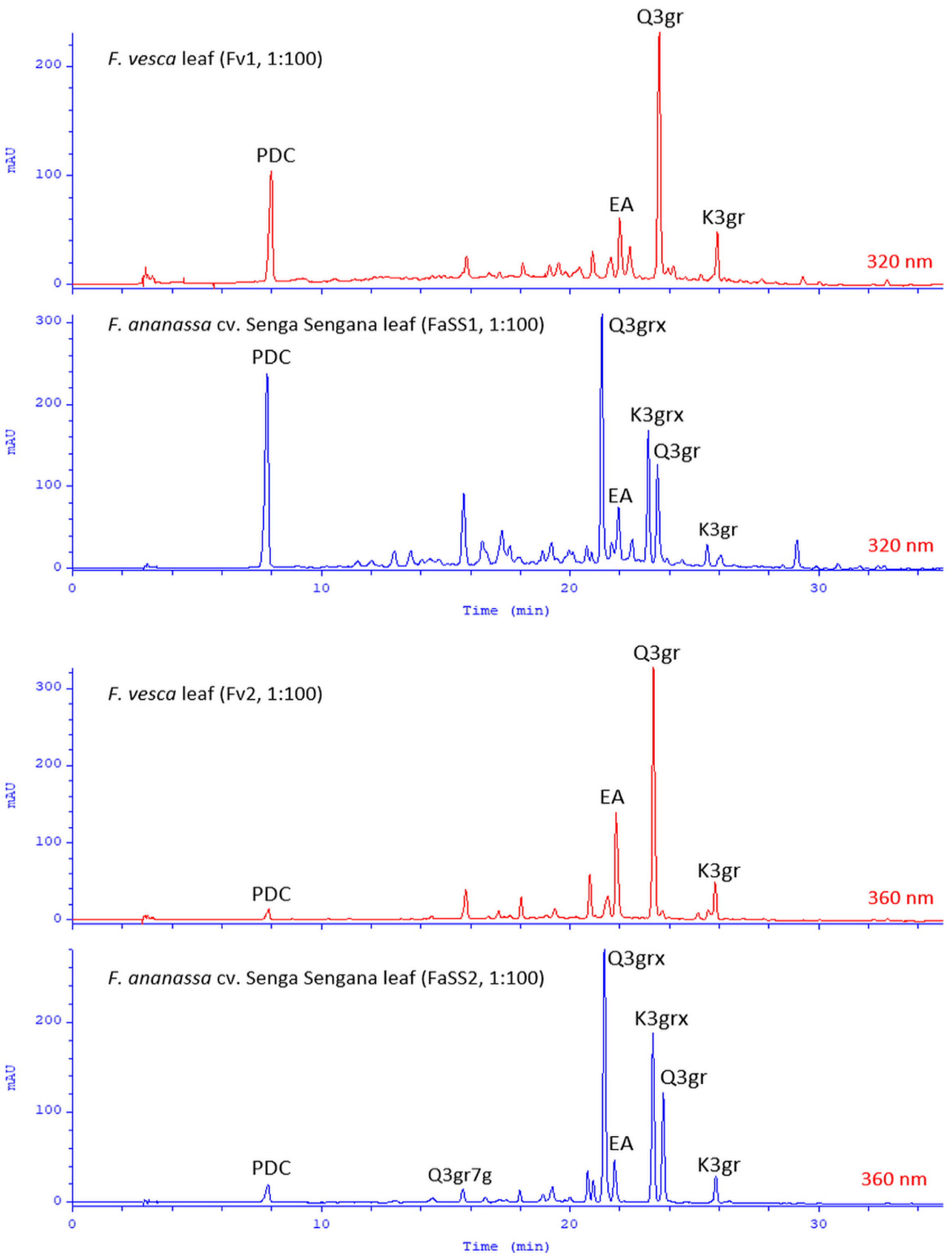
2.5. Chemical Similarity of the Leaves of F. ananassa cv. Senga Sengana and F. vesca

The results obtained from qualitative and quantitative examination of F. ananassa cv. Senga Sengana leaves (FaSS1, FaSS2, FaSS3) were compared with the corresponding data for F. vesca leaves (Fv1 and Fv2). Metabolite profiling showed high similarity between these two species. In woodland strawberry leaves, we observed a comparable PDC content (0.9–1.1% dry matter) and lower levels of ellagitannins and flavonoids. The principal component in both species was agrimoniin (F. vesca 1.48–2.70%). Significant differences were observed in flavonol composition, as the quercetin and kaempferol diglycosides typical for F. ananassa cv. Senga Sengana were absent in F. vesca. Between flavonol derivatives, only 3-O-β-glucuronosides were detected, and their content was slightly higher than in the leaves of the garden strawberry (12, Q3gr, 0.26–0.53%; K3gr, 0.09–0.12%). We also confirmed the occurrence of other minor components. The chemical composition of Fv1 and Fv2 was largely consistent with the scientific literature data [52]. A summary of HPLC chromatograms of F. vesca and F. ananassa cv. Senga Sengana leaf extracts is displayed in Figure 5.

2.6. Antiglycative and Anti-MGO Effects of Flavonols

Flavonols such as quercetin and kaempferol are known for their properties of trapping reactive carbonyl species (RCS), including methylglyoxal (MGO) and glyoxal (GO) [53]. These dicarbonyls originate from the metabolism of simple sugars such as fructose and glucose (from fructolysis and glycolysis in the liver), as well as from lipid peroxidation, and induce carbonyl stress. RCS, regardless of their sources of origin, react with the amino, guanidine and thiol groups of proteins, modifying their structure (post-translational modification) and physiological functions. They also react with lipoproteins, and purine bases in nucleic acids. These reactions result in harmful advanced glycation end products (AGEs) [54]. MGO, produced in excess in the liver, is subsequently secreted into the systemic circulation. Higher plasma MGO levels have been confirmed in hyperglycemic and dyslipidemic subjects and have been linked to metabolic dysfunction, insulin resistance, type 2 diabetes, diabetic retinopathy and nephropathy, non-alcoholic fatty liver (NAFLD), central obesity, atherosclerosis, gout, and other age-related chronic inflammatory diseases such as cardiovascular disease and disorders of the central nervous system. The high MGO concentration in hepatocytes blocks the allosteric binding of AMP to adenosine monophosphate-activated protein kinase (AMPK). Methylglyoxal can modify three arginines in the gamma subunit of AMPK, resulting in its inactivation. AMPK is an enzyme that controls cellular energy, which functions as an energy sensor for metabolic homeostasis and insulin signaling. When AMPK is inhibited, it thereby favors the anabolic processes, including lipogenesis and insulin resistance, which are related to metabolic syndrome and NAFLD [3]. Therefore, strategies that reduce the MGO level through its uptake (neutralization) are considered appropriate for therapeutic or preventive health care. An example of such an intervention is a randomized, double-blind, placebo-controlled, crossover study with quercetin-3-O-β-glucoside (isoquercitrin, 160 mg/day), which observed an 11% reduction in plasma MGO levels in humans [55]. Cardio-metabolic benefits of quercetin in elderly patients with metabolic syndrome were found by Shatylo et al. [56]. The study of Yi et al. [57] summarized quercetin’s therapeutic effects and mechanisms in metabolic diseases.
The two main strawberry quercetin glycosides, Q3gr (12, miquelianin) and Q3grx (11, flagarin), isolated from Senga Sengana leaves, were evaluated for their antiglycation potential in vitro (BSA-MGO model and MGO trapping assays) against known inhibitors such as quercetin, aminoguanidine, and metformin. Metformin is the primary drug used to treat insulin resistance and diabetes type 2. Other quercetin glycosides commonly found in plant materials, including 3-O-β-glucoside (Q3g, isoquercitrin), 4′-O-β-glucoside (Q4′g, spireoside), and 3-O-β-galactoside (Q3ga, hyperoside), were also used in this experiment. Rutin (quercetin-3-O-rutinoside) was examined by us previously [13]. Hyperoside showed the highest antiglycation potential in the assay, and it was comparable to the effect of quercetin (78% vs. 77%) (Table S1). Slightly lower activity (statistically insignificant) was observed for isoquercitrin and miquelianin (12) (75% and 72%)—at the aminoguanidine activity level. Next in line were spireoside (64%), metformin (52%), and 11, quercetin-3-O-[β-xylosyl(1‴→2″)]-β-glucuronoside (45%).
The antiglycation activity of polyphenols is the result of several processes, among which the ability to trap in situ formed RCS, neutralization of reactive oxygen species, as well as reducing and chelating properties, can be considered important. Quercetin and its glycosides are characterized by potent antiradical and reducing effects, and significant antiglycation activity [13]. The type of sugar forming the glycosidic bond and the site of its substitution with the aglycone seem to be essential for this action. Diglycoside 11 (flagarin) was found to be substantially less active compared to the aglycone, but monoglycosides had comparable glycation inhibitory potency (Figure 6), especially those with sugar at the C-3 position (Q3ga = Q ≥ Q3g ≥ Q3gr ≥ Q4′g > Q3grx). It is known that substituting phenolic groups at positions C-5 and C-7 can lose flavonoid MGO trapping potency. Still, position C-4′ (spireoside) only slightly reduces the effect, probably due to the loss of ability to form quinone forms or complexes with metal ions by the catechol group. Interestingly, the 3-O-β-glucuronoside of quercetin (12, miquelianin) retained high glycation inhibitory activity, which gives hope that the antiglycation capacity of flavonol metabolites can be maintained. Indeed, quercetin-3-O-glucuronoside, quercetin-3′-O-sulfate, and isorhamnetin-3-O-glucuronoside (syn. 3′-O-methylquercetin-3-O-glucuronoside) have been identified among the phase II metabolites after oral administration of quercetin glycosides [58,59]. All quercetin monoglycosides better protected BSA from modification compared with metformin. However, it should be noted that these compounds may be less available for different human tissues and organs. After oral intake, plasma concentrations of flavonol metabolites are unfortunately much lower compared to metformin. Nevertheless, they may play an important role in inhibiting glycation in the gut. There is known evidence for the endogenous formation of AGEs in the gastrointestinal tract [60].
In another in vitro test, we examined the ability of quercetin glycosides to trap MGO. The quercetin aglycone was used as the reference substance. All compounds formed adducts with methylglyoxal in vitro. However, di-adducts were noted only for quercetin, hyperoside, spireoside, and miquelianin (Q, Q3ga, Q4′g, and Q3gr) (Table 3). The flavonol A-ring arrangement provides the possibility of MGO addition at two positions, and the resulting adducts take the structure of hemiketal or hemiacetal [61]. Therefore, in the reaction products, we observed several forms of isomeric mono- and di-adducts with different retention times but identical masses, higher by 72 Da or 144 Da, respectively, compared to the precursor. Figure S6 shows the corresponding ions for Q3gr (12) and Q3grx (11) adducts.
Due to the nature of the tannins (their incompatibility with the BSE proteins), this assay was found to be not suitable to measure their suspected antiglycation potential.

3. Discussion

Therapeutic uses of wild strawberry leaf preparations include treating gastrointestinal and urinary tract disorders [8]. Possibly, tannins present in leaves exert an astringent effect on the mucosa and have antimicrobial activity [9,62]. On the other hand, flavonoids are responsible for antioxidant, anti-inflammatory, cytoprotective, and diuretic action [63,64,65,66]. The glycation inhibitory and anti-MGO actions of strawberry flavonols may also be crucial for improving liver metabolism, e.g., quercetin and kaempferol glycosides could protect AMPK against post-translational modification and/or activate AMPK [67]. That effect was described in older scientific sources as ‘improving metabolism’ and ‘blood purification’. Thus, polyphenols identified in the Senga Sengana leaf extracts could contribute to its biological effects. For example, Zhang and coworkers’ findings [68] showed that F. ananassa leaves significantly alleviate cognitive and memory dysfunction in a diabetic animal model. The garden strawberry leaf components exhibited a strong antioxidant effect, reduced the blood glucose of diabetic rats, and improved their cognitive function by regulating the inflammatory response and inhibiting the caspase cascade. Similarly, F. ananassa leaf extract significantly decreased blood glucose, plasma creatinine, urea nitrogen, and renal malondialdehyde in diabetic nephropathy of rats [69]. Kashchenko et al. [70] attributed the hypoglycemic effect of agrimoniin to α-glucosidase inhibition. In experiments by D’Urso et al. [52], an aqueous extract of woodland strawberry leaves possessed direct, endothelium-dependent vasodilatation activity, and its potency was similar to that of an aqueous hawthorn extract. Other researchers reported the inhibitory action of F. vesca leaf extract, and the respective ellagitannin-enriched fraction, in Helicobacter pylori isolates with differential virulence, suggesting the potential of this plant material for the development of new medicines [71]. Furthermore, Juergenliemk et al. [72] confirmed in vitro that quercetin-3-O-glucuronoside (miquelianin) could cross the blood–brain barrier and reach the CNS, which determines its antidepressant effects demonstrated in animal model studies.
Therefore, based on our results and the cited scientific data, we conclude that the compounds identified in the leaves of F. ananassa and F. vesca are bioactive components. Their occurrence explains the biological effects observed in the quoted experiments, which may partially be due to the ability of flavonols to trap MGO and inhibit the harmful glycation of biomolecules. However, the antiglycation properties of flavonol glycosides demonstrated in this study are only preliminary and fraught with the inherent limitations of an in vitro model. Their translation to a therapeutic or prophylactic effect in humans requires further studies, including clinical trials.
In recent years, significant emphasis has been placed on the zero-waste way of life. As a result, different branches of industry, farming, and cultivation are forced to be more productive in a green manner. Some of the prominent examples are mainly connected with cereals or fruit processing, including valorization of by-products [73,74,75,76,77] and technological solutions [78,79]; however, more holistic approaches are found, too [80,81]. Our findings show that waste materials resulting from strawberry production should attract the attention of pharmaceutical companies.

4. Materials and Methods

4.1. Chemicals

LC-MS grade solvents were from Merck (Darmstadt, Germany). Analytical grade diisopropyl ether was purchased from Sigma-Aldrich (St. Louis, MO, USA). Other analytical grade solvents were from POCh (Lublin, Poland). Deuterated solvents were from Armar AG (Döttingen, Switzerland). Methylglyoxal (MGO, 40% in water), bovine serum albumin (BSA, ≥98%), and sodium azide (99%) were purchased from Sigma-Aldrich/Merck; MGO-assay salts (reagent grade) were from Chempur (Piekary Śląskie, Poland).

4.2. Standards

Kaempferol, quercetin, myricetin, taxifolin, catechin, epicatechin, astragalin (K3g, kaempferol-3-O-β-glucoside), kaempferol-3-O-β-glucuronoside (K3gr), nicotiflorin (K3grh, kaempferol-3-O-rutinoside), tiliroside (T, kaempferol-3-O-β-(6″-O-p-coumaroyl) glucoside), isoquercitrin (Q3g, quercetin-3-O-β-glucoside), quercetin-4′-O-β-glucoside (Q4′g, spireoside or spiraeoside), hyperoside (Q3ga, quercetin-3-O-β-galactoside), gallic acid (GA), protocatechuic acid (PA), caffeic acid (CA), and p-coumaric acid (pCuA) with declared purity 99% were from Extrasynthese (Genay, France). Ellagic acid (EA, ≥95%) was purchased from Koch-Light Laboratories (Colnbrook, UK), while rutin (Q3grh, quercetin-3-O-rutinoside), citric acid, quinic acid, and sugar standards were from Merck. Agrimoniin (A) with approximately 97% purity (HPLC-DAD at λ 254 nm) was previously isolated from Potentilla anserina L. [11]. Procyanidins B3 and C2 with approximately 95% purity (HPLC-DAD at λ 254 nm) were isolated from Potentilla erecta (L.) Raeusch. [33]. Isolated compounds analyzed in the antiglycation assay (11, 12) were of at least 95% purity (HPLC-DAD at λ 254 nm, UHPLC-MS).
Stock standard solutions of polyphenols at 1 mg/mL concentration were prepared in methanol, except the ellagic acid (prepared in dimethyl sulfoxide). Working standard solutions (20–800 μg/mL) were prepared by dilution with 50% water-methanol (v/v), then filtered through Durapore 0.22 μm syringe filters (Millipore; Burlington, MA, USA), and stored in the freezer (−20°C).
Metformin hydrochloride (M; Pharmaceutical Secondary Standard) and aminoguanidine hydrochloride (Ag; ≥98%) were purchased from Sigma-Aldrich/Merck. The stock solutions of compounds for in vitro assays (3 mM) were prepared by dissolving reference compound in 5 mL of a suitable solvent and filtered through hydrophilic Millex as above.

4.3. Plant Material

Leaves with petioles of garden strawberry (Fragaria × ananassa Duch. cv. Senga Sengana) were collected from the local plantation in Pasikurowice (district Długołęka, Poland) in July of two consecutive cultivation years (FaSS1, FaSS2; 5 kg each) and from the experimental farm COBORU (Research Centre for Cultivar Testing, Masłowice, Poland) in July of 2020 (FaSS3; 1 kg). The leaves were collected as agricultural waste after fruit harvesting. Before extraction, leaves were air-dried at ambient temperature (22 ± 2 °C), protected from direct sunlight, and crushed. Plant voucher specimens designated as FaSS1, FaSS2, and FaSS3, respectively, are retained in the Herbarium of our Department (Faculty of Pharmacy, Wroclaw Medical University).
Dry leaves of woodland strawberry (Fragaria vesca L.) harvested in July from the Botanical Garden of the University of Wrocław (Poland) were applied as reference material Fv1. A commercial woodland strawberry leaf Fv2 (Flos, Mokrsko, Poland) was used in the comparative analysis.

4.4. Sample Preparation for LC

For the LC analysis, 50% water-methanol extracts (DER 1:100; m/v) were prepared from 0.5 g samples of powdered strawberry leaves and water-methanol mixture (1+1, v/v) in an ultrasonic bath Sonorex Digital 10P (Bandelin, Berlin, Germany). Extraction was performed at an ambient temperature for 15 min [11]. The resulting extracts were transferred to volumetric flasks, brought up to 50 mL with an extraction solvent, and next filtered through Whatman filter papers Grade 1 (Little Chalfont, UK). Fractions, sub-fractions, and isolated compounds were dissolved with water-methanol (1+1, v/v). Samples of all analyzed extracts and solutions were subsequently filtered by syringe filters (Durapore 0.22 μm).

4.5. Isolation of F. ananassa cv. Senga Sengana Leaf Constituents

Dry strawberry leaves of cultivar Senga Sengana (FaSS1, 500 g) were processed chromatographically to yield the following constituents: 1 (235 mg), 2 (386 mg), 3 (420 mg), 4 (4a, 65 mg), 5 (464 mg), 6 (195 mg), 7 (2533 mg), 8 (11 mg), 9 (27 mg), 10 (5817 mg), 11 (330 mg), 12 (216 mg), 13 (48 mg), 14 (25 mg), 15 (17 mg), and 16 (245 mg). Detailed data on the separation process of these components are shown in Figure S1. Retention parameters of isolated compounds together with tentative UHPLC-qTOF-MS/MS identification of non-isolated compounds from FaSS are given in Table 1.

4.6. Identification and Quantification

Structures of compounds 1, 3–5, 7–15 were characterized by spectroscopic evidence (UV-VIS, ESI-MS, 1D- and 2D-NMR) and compared with literature. Compounds 2, 6, and 16 were compared chromatographically (Rf, tR), UV-VIS (λmax), and ESI-MS (m/z) with authentic standards. Compounds 13 and 516 were free from other phenolic impurities (purity ≥ 95%, HPLC-DAD; Section 4.6.2). Compound 4a was accompanied by a dopant of a close derivative 4b. Details of NMR and UV-VIS are available in Supplementary Materials, while HRMS data are in Table 1, along with retention data.

4.6.1. Structure Elucidation Equipment

One-dimensional- and two-dimensional-NMR experiments were performed on Bruker Avance 300 MHz and 500 MHz spectrometers (Bruker BioSpin, Rheinstetten, Germany) using the residual solvent peaks as internal standards. NMR tests for glycosides were conducted in DMSO-d6, ellagitannins in acetone-d6 with D2O (1+1, v/v), and acids and esters in CD3OD. NMR data were analyzed by MestReNova 12 software (Mestrelab Research, Santiago de Compostela, Spain). Direct MS spectra (in negative mode) were recorded in water-methanol (1+1, v/v) on the ESI-qTOF Compact mass spectrometer (Bruker Daltonics, Bremen, Germany). LC-MS-derived spectra were recorded in an appropriate eluent (water-methanol acidified with formic acid) in negative mode. MS data were managed by Data Analysis 4.2 software (Bruker Daltonics). UV-VIS spectra of isolated compounds were measured in water-methanol (1+1, v/v; 0.01–0.03 mM) on a Cecil CE 3021 spectrometer (Cecil Instruments, Cambridge, UK).

4.6.2. Chromatography

TLC was performed on silica gel 60 plates with a fluorescent indicator (Si60 F254, 0.25 mm, 10 × 20 cm; Merck) at ambient temperature. Tested solutions (5–35 μL) were applied as 3–5 mm bands and chromatographed in mobile phases consisting of diisopropyl ether, acetone, formic acid, and water (ellagitannins-E, 4+4+1+1; flavonoids-F, 5+3+1+1; phenolic aids-P, 5+2+2+1; v/v/v/v) [82]. Simple sugars (monosaccharides), liberated during acid hydrolysis, were identified in the mobile phase S-chloroform-methanol-glacial acetic acid-water (8+5+0.2+0.8; v/v/v/v), whereas flavonoid aglycones were identified in the mobile phase X (chloroform-acetone-water, 5+7+8; v/v/v) and Z (chloroform-acetone-formic acid, 8+1+1; v/v/v). Flavonoids were visualized at 254 nm and 366 nm, without and with AlCl3 (2% in methanol) or NP (1% in methanol) spraying. Hydrolyzable tannins and phenolic acids were visualized in VIS using FeCl3 (1% in methanol), whereas monosaccharides with a thymol reagent (0.5 g of thymol in a mixture of 5 mL of concentrated H2SO4 with 95 mL of methanol; heated at 120 °C).
The validated HPLC-DAD method described previously was used to examine the components of strawberry leaf extracts [33]. Separation was achieved on a Hypersil Gold C18 column (250 × 4.6 mm, ∅ 5 μm) with a C18 precolumn (10 × 4.6 mm, ∅ 5 μm) (Thermo Fisher Scientific, Waltham, MA, USA). The linearity of the HPLC-DAD method and quantification of selected polyphenols and PDC was performed based on regression equations determined for the isolated compounds from peak areas and corresponding concentrations.
The Ultimate 3000 system (Thermo Fisher Scientific) with a Kinetex C18 column (150 × 2.1 mm, ∅ 2.6 μm for extracts; 100 × 2.1 mm, ∅ 2.6 μm for MGO derivatives; Phenomenex, Torrance, CA, USA) coupled to the ESI-qTOF Compact mass spectrometer (Bruker Daltonics) was used for qualitative UHPLC-qTOF-MS/MS. Equipment, analysis parameters, and gradient were as reported previously [82](for analysis of extracts), [13](for analysis of MGO adducts).

4.6.3. Acid Hydrolysis

Solutions of isolated hydrolyzable tannins (∼50 mg in 5 mL of 5% H2SO4) were heated (90 °C, reflux, eight hours) according to Tanaka et al. [83]. The hydrolysis products were purified on the octadecyl columns (Isolute C18, 10 g, Biotage, Uppsala, Sweden), then concentrated under reduced pressure and diluted in methanol (5 mL). Phenolic acids and depsides released by hydrolytic degradation were analyzed by TLC (mobile phase P).
Solutions of other polyphenols isolated in small amounts (1 mg in 1 mL 5% H2SO4) were heated at 90 °C in sealed vials for four hours. The hydrolysis products were separated by adding ethyl acetate into water residues [51]. Water layers, concentrated and re-dissolved in 1 mL of methanol, were examined for sugars (TLC, mobile phase S). Organic layers, concentrated and re-dissolved in 1 mL of methanol, were investigated for aglycones (TLC, mobile phases X and Z).

4.6.4. Alkaline Hydrolysis

To initially differentiate the 3-O- and 7-O- position of glycosylation, a small quantity of isolated polyphenol (1 mg) was subjected to alkaline hydrolysis in 1 mL of 0.5% (~0.09 M) aqueous KOH in a boiling water bath for three hours [84].

4.7. Inhibition of Glycation and MGO Trapping In Vitro

4.7.1. Antiglycation Assay in BSA-MGO Model

The formation of advanced glycation end-products (AGEs) was measured following a slightly modified method proposed by Liu et al. [85]. In brief, 21.2 μM bovine serum albumin was incubated with methylglyoxal (MGO) at 0.5 mM in 100 mM PBS at pH 7.4 with 0.02% sodium azide (for prevention of microbial growth). The compounds investigated for inhibition of non-enzymatic glycation were added at a final concentration of 1.5 mM. Then, the reaction solution was incubated at 37 °C and shaken at 50 revolutions per minute for seven days in closed vials away from light. Measurement of the fluorescent intensity of total AGEs after incubation was carried out using a Synergy HTX Multi-Mode Microplate Reader (BioTek Instruments Inc., Winooski, VT, USA) at a wavelength of 360 nm for excitation and 460 nm for emission. Data acquisition was obtained with the Gen5 Software (BioTek Instruments Inc., Winooski, VT, USA). The measurements from three experiments were all performed in triplicate, and the percentage inhibition of AGE formation was calculated using the following equation:
Inhibition of AGE formation = [1 − (Fls/Fl0)] × 100 [%],
where Fl0 is the mean fluorescence intensity of the blank sample and Fls is the mean fluorescence intensity of the sample.

4.7.2. MGO Trapping Assay

Methylglyoxal trapping activity was investigated according to a slightly modified version of the Sang et al. [86] method, as previously described [13]. Briefly, 0.6 mM methylglyoxal was incubated for one hour with 0.2 mM of each compound in 100 mM PBS at pH 7.4 and 37 °C to equate to physiological conditions and shaken at 50 revolutions per minute. The incubation reaction was stopped by adding 2.5 μL of acetic acid and placing the collected samples in an ice-cold water bath. Next, the samples were filtered through hydrophilic syringe filters (Durapore 0.22 μm) and analyzed using UHPLC-qTOF-MS/MS to investigate their ability to form adducts with methylglyoxal. The trapping agent solutions were freshly prepared before each series of experiments was begun, and the pH of the sodium phosphate buffer was determined immediately before use.

4.8. Statistical Analysis

All data are presented as means ± standard deviation (SD). Data were analyzed using the Shapiro–Wilk test to assess normality of distribution, followed by one-way analysis of variance (ANOVA) with Tukey’s multiple comparison test using the GraphPad Prism 6 software; P values equal to or less than 0.05 were considered significant.

5. Conclusions

To summarize, a detailed chemical analysis of F. ananassa cv. Senga Sengana leaves revealed the presence of a broad spectrum of polyphenolic metabolites. Their major components were ellagitannins. Galloyl esters of quinic acid and glucuronosides of flavonols constituted the second group. Proanthocyanidins, flavan-3-ols, and phenolic acid derivatives were minor components. Several new polyphenols including kaempferol and quercetin xylosyl-glucuronosides were also identified.
The occurrence of polyphenols together with 2-pyrone-4,6-dicarboxylic acid at relatively high levels further provides the opportunity to utilize this resource in biotechnological processes to obtain PDC. Moreover, strawberry flavonols have demonstrated the capacity to uptake methylglyoxal and inhibit protein glycation in vitro. Due to that, the leaves of Senga Sengana may become a new raw plant material with therapeutic, pro-health, or cosmetic phytoconstituents.

Supplementary Materials

The following supporting information can be downloaded at: https://www.mdpi.com/article/10.3390/molecules27165293/s1, part A: NMR data of compounds 1, 3, 4a, 5, and 814; part B: Supplementary Figures S1–S9: Scheme of extraction and isolation of individual polyphenols; UV data of compounds 10 vs. 16, 5 vs. GA, 11 vs. 12, 11 vs. 13; MS spectra of MGO adducts with 12 and 13; NMR data for 4a; NMR data for 13; HSQC details for 10; Supplementary Table S1: The antiglycation activity of selected flavonols (incl. 12 and 13) and known inhibitors.

Author Contributions

Conceptualization, I.F.; Data curation, I.F., K.B. and M.W.; Formal analysis, I.F. and M.W.; Funding acquisition, I.F.; Investigation, I.F., K.B. and M.W.; Methodology, I.F. and M.W.; Project administration, I.F.; Resources, I.F.; Supervision, I.F.; Validation, I.F. and K.B.; Visualization, I.F., K.B. and M.W.; Writing—original draft, I.F., K.B. and M.W.; Writing—review and editing, I.F. and M.W. All authors have read and agreed to the published version of the manuscript.

Funding

This research was funded by Wroclaw Medical University, grant no. SUB.D110.21.101. The APC was funded by MDPI.

Institutional Review Board Statement

Not applicable.

Informed Consent Statement

Not applicable.

Data Availability Statement

Not applicable.

Acknowledgments

UHPLC-qTOF-MS/MS and NMR (300 MHz) experiments were performed in the Laboratory of Elemental Analysis and Structural Research, Wroclaw Medical University. NMR (500 MHz) experiments were carried out in the Laboratory of NMR Structural Research, Faculty of Chemistry, University of Wroclaw. The technical assistance of Ruszkiewicz, M., Czapor-Irzabek, H., and Fajfrowski, Ł. is gratefully acknowledged. The authors thank Agnieszka Nowicka, for her assistance in collecting the raw plant material and the donors of plant material for their selflessness.

Conflicts of Interest

The authors declare no conflict of interest.

Sample Availability

Samples of the isolated compounds are available from the authors.

Appendix A. Details on Isolation of F. ananassa cv. Senga Sengana Leaf Constituents

Dry strawberry cv. Senga Sengana leaves (FaSS1, 500 g) were crushed with a hand blender in a water and acetone mixture (1+1, v/v; 6 L) at room temperature. The resulting suspension was filtered by a slit glass (G0) and concentrated under reduced pressure to give the water residue of the aq. acetone extract, FaSS-A (3 L). After acidification with formic acid (up to 0.5%, v/v), FaSS-A was subjected to column chromatography (CC) on octadecyl (J.T. Baker, Phillipsburg, NJ, USA). Components of FaSS-A were eluted using water and methanol in a stepwise gradient manner. The separation of compounds was monitored by TLC Si60 (mobile phases E, F, and P) and HPLC-DAD methods. Eluates with similar compositions were pooled and concentrated to give 13 fractions: FaSS-0 to FaSS-XII. FaSS-0 was eluted with 0.5% formic acid (from the FaSS-A application) and vacuum concentrated. The other fractions were eluted with water-methanol and methanol. Fractions FaSS-I to FaSS-VIII were concentrated under reduced pressure, followed by the SPE method on a C18. This procedure was developed and described previously by Fecka et al. [82]. Fractions FaSS-IX to FaSS-XII were subsequently concentrated under reduced pressure and allowed to crystallize or sediment. Vacuum-dried FaSS-0 was fractionated on a Sephadex LH-20 (Cytiva, Marlborough, MA, USA) by eluting the compounds with methanol. Concentrated FaSS-I was further separated on a Si60 chromatography column (∅ 0.040–0.063 mm; Merck) with methanol–chloroform. Fractions FaSS-II to FaSS-IX were subsequently purified over Sephadex LH-20 with methanol-acetone. Subfractions with similar compositions (TLC, HPLC-DAD) were pooled, concentrated under vacuum (40 °C), and allowed to crystallize or dry. The separated individual compounds were recrystallized from methanol or water–methanol (1+1). The separation steps of F. ananassa cv. Senga Sengana leaf components are shown in Figure S1. The purified compounds were then subjected to structural (spectroscopic) analysis.
Compound 1 (235 mg) was isolated together with 2 (386 mg) from FaSS-0 on Sephadex LH-20 using methanol; 3 (420 mg), 4 (4a, 65 mg) and 5 (464 mg) were obtained from FaSS-I by purification on Si60 with a methanol–chloroform gradient; 6 (195 mg) and 7 (2533 mg) were separated from FaSS-II on Sephadex LH-20 with acetone and methanol–acetone (1+9); 8 (~11 mg) from FaSS-III with methanol–acetone (1+9); 9 (~27 mg) from FaSS-IV with methanol–acetone (1+9); 10 (5817 mg) from FaSS-V with methanol–acetone (2+8); 11 (~330 mg) from FaSS-VI with methanol–acetone (2+8); 12 (216 mg) from FaSS-VII with methanol–acetone (1+9); 13 (~48 mg) from FaSS-VIII with methanol–acetone (2+8); 14 (~25 mg) from FaSS-IX with methanol–acetone (1+9); 15 (~17 mg) from FaSS-X and 16 (245 mg) from FaSS-XI by crystallization with water–methanol (1+1). Retention parameters of isolated compounds together with tentative UHPLC-qTOF-MS/MS identification of non-isolated compounds from FaSS are given in Table 1.
Structures of compounds 1, 35, 715 were characterized by spectroscopic evidence (UV-VIS, ESI-MS, 1D- and 2D-NMR) and compared with literature. Compounds 2, 6, and 16 were compared chromatographically (Rf, tR,), UV-VIS (λmax), and ESI-MS (m/z) with authentic standards. Experimental NMR data are provided in supplementary materials. Compounds 13 and 516 were free from other phenolic impurities (purity ≥ 95%, HPLC-DAD). Compound 4a was accompanied by a dopant of a close derivative.

Appendix B. Details on Identification of F. ananassa cv. Senga Sengana Leaf Constituents

Appendix B.1. Hydrolyzable Tannins

A dimeric ellagitannin (10), isolated as a beige, amorphous powder from FaSS-V and identified as agrimoniin, was the most abundant compound in Senga Sengana leaves. Agrimoniin was previously obtained from Agrimonia pilosa Ledeb. (syn. A. japonica (Miq.) Koidz.) and Potentilla kleiniana Wight et Arnott. [17,18], as well as from F. ananassa Duch. and F. nipponica Makino leaves [29], and berries of woodland and garden strawberry [45]. High-resolution MS1 and MS2 data of 10 with a pseudomolecular ion at m/z 934.0722 [M−2H]−2 (predominant peak; calc. 934.0718 and 1869.1508, [C82H51O52]) are presented in Table 1. Fragmentation of 10 produced ions derived from structures of secondary ellagitannins and low-molecular depsides (i.e., hexahydroxydiphenoyl—HHDP, and dehydrodigalloyl—DHDG; both 302 Da) formed by partial degradation of agrimoniin at m/z: 1567 [M−302(HHDP)−H] (laevigatin B/C/F), 1085 [M−784(pedunculagin)−H] (agrimonic acid A/B lactone), 935 [M−934−H] (potentillin), 897 [M−936(potentillin)−36(2H2O)−H], 783 [M−1086(agrimonic acid A/B lactone)−H] or [M−784(pedunculagin)−302(HHDP)−H] (pedunculagin α/β or laevigatin A lactone), 745 [M−936(potentillin)−152(galloyl)− 36(2H2O)−H], 633 [M−934−302(HHDP)−H] (galloyl-HHDP-glucose), 613 [M−1086(agrimonic acid A/B lactone)−152(gall)−18(H2O)−H], 481 [M−1086(agrimonic acid A/B lactone)−302(HHDP)−H], (HHDP-glucose), 463 [M−1086 (agrimonic acid A/B lactone)−302(HHDP)−18(H2O)−H], 319 [DHDG−18(H2O)−H] (mono-lactone of DHDG), 301 [HHDP−36(2H2O)−H] (dilactone of HHDP, ellagic acid), and 275 [HHDP−18(H2O)−44(CO2)−H] (decarboxylated monolactone of HHDP). The presence of ellagic acid in the acid hydrolysis products of 10 was also confirmed by TLC (mobile phase P).
The 1D- and 2D-NMR experiments of 10 were conducted in acetone-d6 with D2O (1+1, v/v). Since the agrimoniin molecule has four HHDP groups, one DHDG coupler and two α-D-glucopyranoses (α-Glcp), and it is asymmetric [17] in the 1H-NMR spectrum, we observed ten signals typical for these phenolic moieties in an aromatic region and two doublets from anomeric protons of two α-Glcp (Figure S9), as well as other sugar signals. For the DHDG unit, it was δ: 7.31 (1H, d, J = 2 Hz), 7.17 (1H, s); for two HHDP units δ: 6.62, 6.60, 6.53, 6.47, 6.45, 6.31, 6.29, 6.28 (each 1H s); for two α-Glcp anomers δ: 6.49, 6.42 (each 1H, d, J = 3.5 Hz) (α-Glcp1 and α-Glcp2, H-1,1′), and for other α-Glcp protons δ: 5.39, 5.28 (each 1H, t, J = 9.5 Hz, α-Glcp H-3,3′), 5.26, 5.22 (each 1H, dd, α-Glcp H-2,2′), 5.14, 4.86 (each 1H, t, J = 13.2 Hz, α-Glcp H-62,62′), 5.02, 4.93 (1H, t, J = 9.5 Hz, α-Glcp H-4,4′), 4.50, 3.88 (1H, dd, J = 9.5, 6.3 Hz, α-Glcp H-5,5′), 3.75, 3.40 (1H, d, J = 13.2 Hz, α-Glcp H-61,61′). 13C-NMR (and HSQC indirectly) of 10 showed ten ester-forming carboxylic groups δ: 169.78, 169.54, 169.45, 168.98, 168.68, 168.35, 168.29, 165.56, 164.59, 164.19 ppm. The following anomeric α-Glcp carbons were also HSQC-correlated with respective protons of molecule 10: H-1/90.79 ppm and H-1′/90.44 ppm. All carbon signals of agrimoniin are reported in Part A of Supplementary Materials.
Compound 7 isolated from FaSS-II as a beige, amorphous powder with a pseudomolecular ion at m/z 783.0676 (calc. 783.0686, [C34H23O22]) and fragmentation typical for hexahydroxydiphenoyl esters (481, 301, 275) was tentatively identified as a monomeric ellagitannin named pedunculagin. 1H-NMR analysis of 7 confirmed this assumption [16].
Compound 3 and 5 (from FaSS-I) with identical pseudomolecular ions at m/z 343.0669 [M−H] (predominant, calc. 343.0671, [C14H15O10]) produced fragments deriving from both quinic (191 [QA−H], 173 [QA−18(H2O)−H]) and gallic acids (169 [GA−H], 125 [GA−44(CO2)−H]). A galloyl moiety was also detected after acid hydrolysis of 3 and 5 (TLC, mobile phase P). Those monogalloylquinic acid regioisomers showed UV-VIS spectra with the 2nd absorption maximum at 273 nm, typical for galloyl esters. The NMR data of 3 and 5 are summarized in Supplementary Materials. Monogalloylquinic acids 3 and 5 (classified in gallotannins) were therefore identified as 3- and 5-O-galloylquinic acid, respectively [14,15].

Appendix B.2. Proanthocyanidins and Flavan-3-Ols

Condensed tannins were minor F. ananassa cv. Senga Sengana components. Several B-type proanthocyanidins (five dimers, three trimers, and one tetramer) were detected together with monomeric catechin by UHPLC-qTOF-MS/MS (m/z 289.0719, 577.1346, 865.1981, 1153.2610). Catechin (6) was also isolated as a fine white powder from fraction FaSS-II. Chromatographic and spectroscopic data (Rf, tR, UV-VIS, ESI-MS) of 6 were consistent with the authentic standard of this compound. Procyanidins B3 and C2 as well as other oligomeric flavan-3-ol derivatives of both catechin and epicatechin were recognized based on UV-VIS profiles and MS fragmentations. Three distinct fragmentation pathways were observed for these proanthocyanidins: loss of neutral molecules by HRF (heterocyclic ring fission, −126 Da), RDA (retro-Diels-Alder fission, −152 Da), and QM (quinone methide fission, Δ 2H) [87,88]. In particular, fragment ions released during C-C bond fission by QM were diagnostically relevant in the analysis of B-type proanthocyanidins, e.g., for procyanidin B3 these ions were at 287 and 289, for procyanidin C2 at 575, 577, 287 and 289 (Table 1). Catechin and procyanidins B3 and B6 were formerly isolated from F. ananassa cv. Reikov roots [26].

Appendix B.3. Flavonoids

Three diglycosides, i.e., 9 (from FaSS-IV), 11 (from FaSS-VI), and 13 (from FaSS-VII), as well as four monoglycosides, i.e., 8 (FaSS-III), 12 (FaSS-VII), 14 (FaSS-IX) and 15 (FaSS-X), were isolated from FaSS-A. Glucuronic acid, glucose, and xylose were detected in their glycone parts after acid hydrolysis. The sugar-aglycone connections were based on HMBC/NOESY, as well as by other experiments (Section 4.6). Some of the noted flavonoids, including flavonol diglycosides (MW 594, 610, and 640 Da), were described previously in leaves of F. ananassa cv. Polka [27], in flowers of cv. Jonsok [28], and in pseudo-fruits of the Japanese cultivar Tochiotome [21]. Nevertheless, their complete structure has not been established or finely defined.
Compound 9 (yellow crystalline powder) exhibited a pseudomolecular ion at m/z 639.1200 [M−H] (calc. 639.1203 for [C27H27O18]) that is relevant to hexoside-hexuronide of quercetin-like aglycone (ions at 301, 179 and 151). The MS2 spectrum revealed the presence of a fragment ion at m/z 463 [M−176−H] corresponding to partially degraded diglycoside after the loss of hexuronic acid and suggested that sugars must be attached independently. Both glucose and glucuronic acid were released by acidic hydrolysis, confirmed by TLC (mobile phase S). The 1D and 2D-NMR experiments on 9 were conducted in DMSO-d6. All data were similar to those presented by Felser and Schimmer [20]. Briefly, the typical quercetin pattern of proton signals was not affected by glycosylation in flavonol ring B. A glucose (β-Glcp) moiety was connected at position C-7 of aglycone, which was proved by NOESY (H-1‴↔H-8). The glucuronoside moiety was attached at position C-3 (H-1 of β-GlcAp showed HMBC correlation with C-3). Finally, compound 9 was labelled as quercetin-3-O-β-glucuronoside-7-O-β-glucoside.
The MS spectrum of compound 8 (beige powder) with ions at m/z 465.1035 [M−H] (calc. 465.1039, [C21H21O12]), 303 [M−162−H] and 285 [303−18−H] was relevant to hexoside of dihydroquercetin (taxifolin). The loss of a water molecule from the aglycone during fragmentation of 8 demonstrated unambiguously its flavanonol structure and indicated the likely sugar substitution position at C-3. The NMR analysis was conducted in CD3OD to compare the results with the reference. All spectroscopic data of 8 were clearly similar to those presented by Sakushima et al. [19] and different from those of Pan and Lundgren [47], but the spectrum revealed a number of overlapping isomer signals. The typical dihydroquercetin pattern of proton signals was not affected by glycosylation in flavonoid rings A and B. Glycosylation at position C-3 was based on shifts of H-2 and H-3: 5.24 (d, J = 9.7 Hz) and 4.93 ppm (d, J = 9.8 Hz), in comparison to unsubstituted 2,3-dihydroquercetin. The absolute configuration at C-2 and C-3 of the main compound in the mixture was provisionally assigned by comparison with Sakushima et al. [19] to be 3-O-β-glucoside of (2R,3R)-taxifolin (dihydroquercetin). That compound was also found in Tochiotome strawberries by Abe et al. [21]. Other taxifolin pentosides and hexosides were also observed in LC-MS as minor components (Table 1). For example, taxifolin-3-O-α-arabinoside was isolated previously from roots of F. ananassa cv. Reikov by Ishimaru et al. [26].
Based on spectroscopic evidence, flavonoids 12, 14 and 15 were identified as quercetin-3-O-β-glucuronoside (syn. quercetin-3-O-β-glucuronide or miquelianin, m/z 477.0672), isorhamnetin-3-O-β-glucuronoside (3′-O-methylquercetin-3-O-β-glucuronoside, 491.0806), and tiliroside (kaempferol-3-O-β-(6″-O-p-coumaroyl) glucoside, 593.1299; a pair of cis/trans isomers), respectively [22,23,24,38,39,40,41]. Using UHPLC-qTOF-MS/MS, the following flavonoids were additionally detected: quercetin-3-O-β-(glucosyl)glucuronoside (tentatively, based on MS and MS2)—m/z 639.1200 [M−H], quercetin-3-O-β-glucoside (isoquercitrin, confirmed with standard)—463.0878 [M−H], kaempferol-3-O-β-glucuronoside (confirmed with standard)—461.0724 [M−H] and kaempferol-3-O-β-glucoside (astragalin, confirmed with standard)—447.0929 [M−H]. A different fragmentation pattern than for quercetin-3-O-β-glucuronoside-7-O-β-glucoside characterized quercetin-3-O-β-(glucosyl)glucuronoside. Its glycone moiety, structurally similar to 3-O-[β-xylosyl(1‴→2″)]-β-glucuronosides of quercetin and kaempferol, was uncoupled as a whole disaccharide chain [M−338−H], in applied conditions of ESI-qTOF-MS/MS.

Appendix B.4. Phenolic and Carboxylic Acids

UHPLC-qTOF-MS/MS analysis of F. ananassa cv. Senga Sengana leaf extracts showed intense peaks of several phenolic acid derivatives, as well as carboxylic acids. Compound 1 was isolated from FaSS-0 as a white crystalline powder with λmax at 315 nm, and pseudomolecular ions at m/z 182.9934 [M−H] (predominant, calc. 182.9935, [C7H3O6]) and 366.9940 [2M−H]. Its fragmentation-derived ions suggested a subsequent neutral loss of carbon dioxide and carbon oxide: 139 [M−44/CO2−H] and 111 [M−44/CO2−28/CO−H]. The 1H- and 13C-NMR (in CD3OD) data of 1 were consistent with results published by Wilkes and Glasl [25]. Thus, 1 was identified as 2-pyrone-4,6-dicarboxylic acid (PDC).

References

  1. Koch, W. Dietary polyphenols—Important non-nutrients in the prevention of chronic noncommunicable diseases. A systematic review. Nutrients 2019, 11, 1039. [Google Scholar] [CrossRef] [PubMed]
  2. Zuo, L.; Prather, E.R.; Stetskiv, M.; Garrison, D.E.; Meade, J.R.; Peace, T.I.; Zhou, T. Inflammaging and oxidative stress in human diseases: From molecular mechanisms to novel treatments. Int. J. Mol. Sci. 2019, 20, 4472. [Google Scholar] [CrossRef] [PubMed]
  3. Mortera, R.R.; Bains, Y.; Gugliucci, A. Fructose at the crossroads of the metabolic syndrome and obesity epidemics. Front. Biosci. Landmark 2019, 24, 186–211. [Google Scholar]
  4. Yang, C.S.; Wang, H.; Sheridan, Z.P. Studies on prevention of obesity, metabolic syndrome, diabetes, cardiovascular diseases and cancer by tea. J. Food Drug Anal. 2018, 26, 1–13. [Google Scholar] [CrossRef]
  5. Liu, J.; He, Z.; Ma, N.; Chen, Z.Y. Beneficial effects of dietary polyphenols on high-fat diet-induced obesity linking with modulation of gut microbiota. J. Agric. Food Chem. 2020, 68, 33–47. [Google Scholar] [CrossRef] [PubMed]
  6. Catalkaya, G.; Venema, K.; Lucini, L.; Rocchetti, G.; Delmas, D.; Daglia, M.; De Filippis, A.; Xiao, H.; Quiles, J.L.; Xiao, J.; et al. Interaction of dietary polyphenols and gut microbiota: Microbial metabolism of polyphenols, influence on the gut microbiota, and implications on host health. Food Front. 2020, 1, 109–133. [Google Scholar] [CrossRef]
  7. García-Villalba, R.; Vissenaekens, H.; Pitart, J.; Romo-Vaquero, M.; Espín, J.C.; Grootaert, C.; Selma, M.V.; Raes, K.; Smagghe, G.; Possemiers, S.; et al. Gastrointestinal simulation model TWIN-SHIME shows differences between human urolithin-metabotypes in gut microbiota composition, pomegranate polyphenol metabolism, and transport along the intestinal tract. J. Agric. Food Chem. 2017, 65, 5480–5493. [Google Scholar] [CrossRef]
  8. Widy-Tyszkiewicz, E. Assessment Report on Fragaria Vesca L., Fragaria Moschata Weston, Fragaria Viridis Weston and Fragaria × Ananassa (Weston) Duchesne ex Rozier, Folium. 2018. Available online: https://www.ema.europa.eu/en/documents/herbal-report/final-assessment-report-fragaria-vesca-l-fragaria-moschata-weston-fragaria-viridis-weston-fragaria-x_en.pdf (accessed on 28 July 2022).
  9. Blaschek, W.; Ebel, S.; Hackental, E.; Holzgrabe, U.; Keller, K.; Reichling, J.; Schulz, J. (Eds.) Fragariae folium. In Hagers Handbuch der Drogen und Arzneistoffe; HagerRom; Springer Medizin Verlag: Heidelberg, Germany, 2006; ISBN 978-3540282327. [Google Scholar]
  10. Wichtl, M.; Blaschek, W. (Eds.) Fragariae folium. In Teedrogen und Phytopharmaka: Ein Handbuch für die Praxis; Wissenschaftliche Verlagsgesellschaft: Stuttgart, Germany, 2016; pp. 262–263. ISBN 978-3804730687. [Google Scholar]
  11. Fecka, I. Development of chromatographic methods for determination of agrimoniin and related polyphenols in pharmaceutical products. J. AOAC Int. 2009, 92, 410–418. [Google Scholar] [CrossRef]
  12. Tridge Report: Strawberry Production. Available online: https://www.tridge.com/intelligences/stawberry/production (accessed on 28 July 2022).
  13. Bednarska, K.; Fecka, I. Potential of vasoprotectives to inhibit non-enzymatic protein glycation, and reactive carbonyl and oxygen species uptake. Int. J. Mol. Sci. 2021, 22, 10026. [Google Scholar] [CrossRef]
  14. Clifford, M.N.; Stoupi, S.; Kuhnert, N. Profiling and characterization by LC-MSn of the galloylquinic acids of green tea, tara tannin, and tannic acid. J. Agric. Food Chem. 2007, 55, 2797–2807. [Google Scholar] [CrossRef]
  15. Nishimura, H.; Nonaka, G.I.; Nishioka, I. Seven quinic acid gallates from Quercus stenophylla. Phytochemistry 1984, 23, 2621–2623. [Google Scholar] [CrossRef]
  16. Feldman, K.S.; Smith, R.S. Ellagitannin chemistry. First total synthesis of the 2,3- and 4,6-coupled ellagitannin pedunculagin. J. Org. Chem. 1996, 61, 2606–2612. [Google Scholar] [CrossRef]
  17. Okuda, T.; Yoshida, T.; Kuwahara, M.; Memon, M.U.; Shingu, T. Agrimoniin and potentillin, an ellagitannin dimer and monomer having an α-glucose core. J. Chem. Soc. Chem. Commun. 1982, 3, 162–163. [Google Scholar] [CrossRef]
  18. Okuda, T.; Yoshida, T.; Kuwahara, M.; Memon, M.U.; Shingu, T. Tannins of rosaceous medicinal plants. I. Structures of potentillin, agrimonic acids A and B, and agrimoniin, a dimeric ellagitannin. Chem. Pharm. Bull. (Tokyo) 1984, 32, 2165–2173. [Google Scholar] [CrossRef]
  19. Sakushima, A.; Ohno, K.; Coskun, M.; Seki, K.I.; Ohkura, K. Separation and identification of taxifolin 3-O-glucoside isomers from Chamaecyparis obtusa (Cupressaceae). Nat. Prod. Lett. 2002, 16, 383–387. [Google Scholar] [CrossRef]
  20. Felser, C.; Schimmer, O. Flavonoid glycosides from Alchemilla speciosa. Planta Med. 1999, 65, 668–670. [Google Scholar] [CrossRef]
  21. Abe, T.; Yoshida, S.; Kawabata, T.; Takano, F.; Ohta, T. Study of inhibitory effect of polyphenols from Fragaria ananassa on fat accumulation in rat white adipocyte. JJCAM 2018, 15, 1–7. [Google Scholar] [CrossRef]
  22. Ossipov, V.; Nurmi, K.; Loponen, J.; Prokopiev, N.; Haukioja, E.; Pihlaja, K. HPLC isolation and identification of flavonoids from white birch Betula pubescens leaves. Biochem. Syst. Ecol. 1995, 23, 213–222. [Google Scholar] [CrossRef]
  23. Im, S.H.; Wang, Z.; Lim, S.S.; Lee, O.-H.; Kang, I.-J. Bioactivity-guided isolation and identification of anti-adipogenic compounds from Sanguisorba officinalis. Pharm. Biol. 2017, 55, 2057–2064. [Google Scholar] [CrossRef]
  24. Timmers, M.; Urban, S. On-line (HPLC-NMR) and off-line phytochemical profiling of the Australian plant, Lasiopetalum macrophyllum. Nat. Prod. Commun. 2011, 6, 1605–1616. [Google Scholar] [CrossRef]
  25. Wilkes, S.; Glasl, H. Isolation, characterization, and systematic significance of 2-pyrone-4,6-dicarboxylic acid in Rosaceae. Phytochemistry 2001, 58, 441–449. [Google Scholar] [CrossRef]
  26. Ishimaru, K.; Omoto, T.; Asai, I.; Ezaki, K.; Shimomura, K. Taxifolin 3-arabinoside from Fragaria × ananassa. Phytochemistry 1995, 40, 345–347. [Google Scholar] [CrossRef]
  27. Kårlund, A.; Salminen, J.P.; Koskinen, P.; Ahern, J.R.; Karonen, M.; Tiilikkala, K.; Karjalainen, R.O. Polyphenols in strawberry (Fragaria × ananassa) leaves induced by plant activators. J. Agric. Food Chem. 2014, 62, 4592–4600. [Google Scholar] [CrossRef]
  28. Hanhineva, K.; Rogachev, I.; Kokko, H.; Mintz-Oron, S.; Venger, I.; Kärenlampi, S.; Aharoni, A. Non-targeted analysis of spatial metabolite composition in strawberry (Fragaria × ananassa) flowers. Phytochemistry 2008, 69, 2463–2481. [Google Scholar] [CrossRef]
  29. Okuda, T.; Yoshida, T.; Hatano, T.; Iwasaki, M.; Kubo, M.; Orime, T.; Yoshizaki, M.; Naruhashi, N. Hydrolysable tannins as chemotaxonomic markers in the Rosaceae. Phytochemistry 1992, 31, 3091–3096. [Google Scholar] [CrossRef]
  30. Josuttis, M.; Verrall, S.; Stewart, D.; Krüger, E.; McDougall, G.J. Genetic and environmental effects on tannin composition in strawberry (Fragaria × ananassa) cultivars grown in different European locations. J. Agric. Food Chem. 2013, 61, 790–800. [Google Scholar] [CrossRef]
  31. Hatano, T.; Yazaki, K.; Okonogi, A.; Okuda, T. Tannins of Stachyurus species. II. Praecoxins A, B, C and D, four new hydrolyzable tannins from Stachyurus praecox leaves. Chem. Pharm. Bull. (Tokyo) 1991, 39, 1689–1693. [Google Scholar] [CrossRef]
  32. Karlińska, E.; Pecio, Ł.; Macierzyński, J.; Stochmal, A.; Kosmala, M. Structural elucidation of the ellagitannin with a molecular weight of 2038 isolated from strawberry fruit (Fragaria ananassa Duch.) and named fragariin A. Food Chem. 2019, 296, 109–115. [Google Scholar] [CrossRef] [PubMed]
  33. Fecka, I.; Kucharska, A.Z.; Kowalczyk, A. Quantification of tannins and related polyphenols in commercial products of tormentil (Potentilla tormentilla). Phytochem. Anal. 2015, 26, 353–366. [Google Scholar] [CrossRef] [PubMed]
  34. Yoshida, T.; Tanaka, K.; Xin-min, C.; Okuda, T. Dimeric ellagitannins, laevigatins E, F and G, from Rosa laevigata. Phytochemistry 1989, 28, 2451–2454. [Google Scholar] [CrossRef]
  35. Yoshida, T.; Tanaka, K.; Chen, X.-M.; Okuda, T. Tannis of rosaceous medicinal plants. V. Hydrolyzable tannis with dehydrodigalloyl group from Rosa laevigata Michx. Chem. Pharm. Bull. (Tokyo) 1989, 37, 920–924. [Google Scholar] [CrossRef]
  36. Aaby, K.; Mazur, S.; Nes, A.; Skrede, G. Phenolic compounds in strawberry (Fragaria × ananassa Duch.) fruits: Composition in 27 cultivars and changes during ripening. Food Chem. 2012, 132, 86–97. [Google Scholar] [CrossRef]
  37. Yoshida, T.; Jin, Z.-X.; Okuda, T. Hydrolysable tannin oligomers from Rosa davurica. Phytochemistry 1991, 30, 2747–2752. [Google Scholar] [CrossRef]
  38. Weber, N.; Veberic, R.; Mikulic-Petkovsek, M.; Stampar, F.; Koron, D.; Munda, A.; Jakopic, J. Metabolite accumulation in strawberry (Fragaria × ananassa Duch.) fruits and runners in response to Colletotrichum nymphaeae infection. Physiol. Mol. Plant Pathol. 2015, 92, 119–129. [Google Scholar] [CrossRef]
  39. Itoh, T.; Ninomiya, M.; Yasuda, M.; Koshikawa, K.; Deyashiki, Y.; Nozawa, Y.; Akao, Y.; Koketsu, M. Inhibitory effects of flavonoids isolated from Fragaria ananassa Duch. on IgE-mediated degranulation in rat basophilic leukemia RBL-2H3. Bioorg. Med. Chem. 2009, 17, 5374–5379. [Google Scholar] [CrossRef]
  40. Ninomiya, M.; Itoh, T.; Ishikawa, S.; Saiki, M.; Narumiya, K.; Yasuda, M.; Koshikawa, K.; Nozawa, Y.; Koketsu, M. Phenolic constituents isolated from Fragaria ananassa Duch. inhibit antigen-stimulated degranulation through direct inhibition of spleen tyrosine kinase activation. Bioorg. Med. Chem. 2010, 18, 5932–5937. [Google Scholar] [CrossRef]
  41. Tsukamoto, S.; Tomise, K.; Aburatani, M.; Onuki, H.; Hirorta, H.; Ishiharajima, E.; Ohta, T. Isolation of cytochrome p450 inhibitors from strawberry fruit, Fragaria ananassa. J. Nat. Prod. 2004, 67, 1839–1841. [Google Scholar] [CrossRef]
  42. Hanhineva, K.; Soininen, P.; Anttonen, M.J.; Kokko, H.; Rogachev, I.; Aharoni, A.; Laatikainen, R.; Kärenlampi, S. NMR and UPLC-qTOF-MS/MS characterisation of novel phenylethanol derivatives of phenylpropanoid glucosides from the leaves of strawberry (Fragaria × ananassa cv. Jonsok). Phytochem. Anal. 2009, 20, 353–364. [Google Scholar] [CrossRef]
  43. Lunkenbein, S.; Bellido, M.; Aharoni, A.; Salentijn, E.M.J.; Kaldenhoff, R.; Coiner, H.A.; Munoz-Blanco, J.; Schwab, W. Cinnamate metabolism in ripening fruit. Characterization of a UDP-glucose: Cinnamate glucosyltransferase from strawberry. Plant Physiol. 2006, 140, 1047–1058. [Google Scholar] [CrossRef]
  44. Gasperotti, M.; Masuero, D.; Guella, G.; Palmieri, L.; Martinatti, P.; Pojer, E.; Mattivi, F.; Vrhovsek, U. Evolution of ellagitannin content and profile during fruit ripening in Fragaria spp. J. Agric. Food Chem. 2013, 61, 8597–8607. [Google Scholar] [CrossRef]
  45. Vrhovsek, U.; Guella, G.; Gasperotti, M.; Pojer, E.; Zancato, M.; Mattivi, F. Clarifying the identity of the main ellagitannin in the fruit of the strawberry, Fragaria vesca and Fragaria ananassa Duch. J. Agric. Food Chem. 2012, 60, 2507–2516. [Google Scholar] [CrossRef] [PubMed]
  46. Karlińska, E.; Masny, A.; Cieślak, M.; Macierzyński, J.; Pecio, Ł.; Stochmal, A.; Kosmala, M. Ellagitannins in roots, leaves, and fruits of strawberry (Fragaria × ananassa Duch.) vary with developmental stage and cultivar. Sci. Hortic. 2021, 275, 109665. [Google Scholar] [CrossRef]
  47. Pan, H.; Lundgren, L.N. Phenolic extractives from root bark of Picea abies. Phytochemistry 1995, 39, 1423–1428. [Google Scholar] [CrossRef]
  48. Yang, D.; Xie, H.; Jiang, Y.; Wei, X. Phenolics from strawberry cv. Falandi and their antioxidant and α-glucosidase inhibitory activities. Food Chem. 2016, 194, 857–863. [Google Scholar] [CrossRef]
  49. Li, W.; Shen, X.; Wang, J.; Sun, X.; Yuan, Q. Engineering microorganisms for the biosynthesis of dicarboxylic acids. Biotechnol. Adv. 2021, 48, 107710. [Google Scholar] [CrossRef]
  50. Yamagaki, T.; Watanabe, T. Hydrogen radical removal causes complex overlapping isotope patterns of aromatic carboxylic acids in negative-ion matrix-assisted laser desorption/ionization mass spectrometry. Mass Spectrom. 2012, 1, A0005. [Google Scholar] [CrossRef]
  51. Markham, K.R. Techniques of Flavonoids Identification; Academic Press: London, UK, 1982; ISBN 0-12-472680-1. [Google Scholar]
  52. D’Urso, G.; Pizza, C.; Piacente, S.; Montoro, P. Combination of LC-MS based metabolomics and antioxidant activity for evaluation of bioactive compounds in Fragaria vesca leaves from Italy. J. Pharm. Biomed. Anal. 2018, 150, 233–240. [Google Scholar] [CrossRef]
  53. Li, X.; Zheng, T.; Sang, S.; Lv, L. Quercetin inhibits advanced glycation end product formation by trapping methylglyoxal and glyoxal. J. Agric. Food Chem. 2014, 62, 12152–12158. [Google Scholar] [CrossRef]
  54. Brings, S.; Fleming, T.; Freichel, M.; Muckenthaler, M.; Herzig, S.; Nawroth, P. Dicarbonyls and advanced glycation end-products in the development of diabetic complications and targets for intervention. Int. J. Mol. Sci. 2017, 18, 984. [Google Scholar] [CrossRef]
  55. Van den Eynde, M.D.G.; Geleijnse, J.M.; Scheijen, J.L.J.M.; Hanssen, N.M.J.; Dower, J.I.; Afman, L.A.; Stehouwer, C.D.A.; Hollman, P.C.H.; Schalkwijk, C.G. Quercetin, but not epicatechin, decreases plasma concentrations of methylglyoxal in adults in a randomized, double-blind, placebo-controlled, crossover trial with pure flavonoids. J. Nutr. 2018, 148, 1911–1916. [Google Scholar] [CrossRef]
  56. Shatylo, V.; Antoniuk-Shcheglova, I.; Naskalova, S.; Bondarenko, O.; Havalko, A.; Krasnienkov, D.; Zabuga, O.; Kukharskyy, V.; Guryanov, V.; Vaiserman, A. Cardio-metabolic benefits of quercetin in elderly patients with metabolic syndrome. PharmaNutrition 2021, 15, 100250. [Google Scholar] [CrossRef]
  57. Yi, H.; Peng, H.; Wu, X.; Xu, X.; Kuang, T.; Zhang, J.; Du, L.; Fan, G. The therapeutic effects and mechanisms of quercetin on metabolic diseases: Pharmacological data and clinical evidence. Oxid. Med. Cell. Longev. 2021, 2021, 6678662. [Google Scholar] [CrossRef]
  58. Hai, Y.; Zhang, Y.; Liang, Y.; Ma, X.; Qi, X.; Xiao, J.; Xue, W.; Luo, Y.; Yue, T. Advance on the absorption, metabolism, and efficacy exertion of quercetin and its important derivatives. Food Front. 2020, 1, 420–434. [Google Scholar] [CrossRef]
  59. Almeida, A.F.; Borge, G.I.A.; Piskula, M.; Tudose, A.; Tudoreanu, L.; Valentová, K.; Williamson, G.; Santos, C.N. Bioavailability of quercetin in humans with a focus on interindividual variation. Compr. Rev. Food Sci. Food Saf. 2018, 17, 714–731. [Google Scholar] [CrossRef]
  60. Van der Lugt, T.; Opperhuizen, A.; Bast, A.; Vrolijk, M.F. Dietary advanced glycation endproducts and the gastrointestinal tract. Nutrients 2020, 12, 2814. [Google Scholar] [CrossRef]
  61. Bhuiyan, M.N.I.; Mitsuhashi, S.; Sigetomi, K.; Ubukata, M. Quercetin inhibits advanced glycation end product formation via chelating metal ions, trapping methylglyoxal, and trapping reactive oxygen species. Biosci. Biotechnol. Biochem. 2017, 81, 882–890. [Google Scholar] [CrossRef]
  62. Van Wyk, B.-E.; Wink, M. Fragaria vesca wild strawberry. In Medicinal Plants of the World: An Illustrated Scientific Guide to Important Medicinal Plants and Their Uses; Briza Publications: Pretoria, South Africa, 2004; p. 146. ISBN 1-875093-44-3. [Google Scholar]
  63. Marunaka, Y. Actions of quercetin, a flavonoid, on ion transporters: Its physiological roles. Ann. N. Y. Acad. Sci. 2017, 1398, 142–151. [Google Scholar] [CrossRef]
  64. Vargas, F.; Romecín, P.; García-Guillén, A.I.; Wangesteen, R.; Vargas-Tendero, P.; Paredes, M.D.; Atucha, N.M.; García-Estañ, J. Flavonoids in kidney health and disease. Front. Physiol. 2018, 9, 394. [Google Scholar] [CrossRef]
  65. Wang, X.; Cao, Y.; Chen, S.; Lin, J.; Bian, J.; Huang, D. Anti-inflammation activity of flavones and their structure–activity relationship. J. Agric. Food Chem. 2021, 69, 7285–7302. [Google Scholar] [CrossRef]
  66. Dhanya, R. Quercetin for managing type 2 diabetes and its complications, an insight into multitarget therapy. Biomed. Pharmacother. 2022, 146, 112560. [Google Scholar] [CrossRef]
  67. Yong, Y.; Shin, S.Y.; Jung, Y.; Jung, H.; Ahn, S.; Chong, Y.; Lim, Y. Flavonoids activating adenosine monophosphate-activated protein kinase. J. Korean Soc. Appl. Biol. Chem. 2015, 58, 13–19. [Google Scholar] [CrossRef]
  68. Zhang, L.; Ma, Q.; Zhou, Y. Strawberry leaf extract treatment alleviates cognitive impairment by activating Nrf2/HO-1 signaling in rats with streptozotocin-induced diabetes. Front. Aging Neurosci. 2020, 12, 201. [Google Scholar] [CrossRef]
  69. Ibrahim, D.S.; Abd El-Maksoud, M.A.E. Effect of strawberry (Fragaria × ananassa) leaf extract on diabetic nephropathy in rats. Int. J. Exp. Pathol. 2015, 96, 87–93. [Google Scholar] [CrossRef]
  70. Kashchenko, N.; Chirikova, N.; Olennikov, D. Agrimoniin, an active ellagitannin from Comarum palustre herb with anti-α-glucosidase and antidiabetic potential in streptozotocin-induced diabetic rats. Molecules 2017, 22, 73. [Google Scholar] [CrossRef]
  71. Cardoso, O.; Donato, M.M.; Luxo, C.; Almeida, N.; Liberal, J.; Figueirinha, A.; Batista, M.T. Anti-Helicobacter pylori potential of Agrimonia eupatoria L. and Fragaria vesca. J. Funct. Foods 2018, 44, 299–303. [Google Scholar] [CrossRef]
  72. Juergenliemk, G.; Boje, K.; Huewel, S.; Lohmann, C.; Galla, H.-J.; Nahrstedt, A. In vitro studies indicate that miquelianin (quercetin 3-O-β-D-glucuronopyranoside) is able to reach the CNS from the small intestine. Planta Med. 2003, 69, 1013–1017. [Google Scholar]
  73. Albergamo, A.; Costa, R.; Bartolomeo, G.; Rando, R.; Vadalà, R.; Nava, V.; Gervasi, T.; Toscano, G.; Germanò, M.P.; D’Angelo, V.; et al. Grape water: Reclaim and valorization of a by-product from the industrial cryoconcentration of grape (Vitis vinifera) must. J. Sci. Food Agric. 2020, 100, 2971–2981. [Google Scholar] [CrossRef]
  74. Mohammed, M.A.; Ibrahim, B.M.M.; Abdel-Latif, Y.; Hassan, A.H.; El Raey, M.A.; Hassan, E.M.; El-Gengaihi, S.E. Pharmacological and metabolomic profiles of Musa acuminata wastes as a new potential source of anti-ulcerative colitis agents. Sci. Rep. 2022, 12, 10595. [Google Scholar] [CrossRef]
  75. Petrović, M.; Jovanović, M.; Lević, S.; Nedović, V.; Mitić-Ćulafić, D.; Živković Semren, T.; Veljović, S. Valorization potential of Plantago major L. solid waste remaining after industrial tincture production: Insight into the chemical composition and bioactive properties. Waste Biomass Valorization 2022, 13, 1639–1651. [Google Scholar] [CrossRef]
  76. Slavov, A.; Yantcheva, N.; Vasileva, I. Chamomile wastes (Matricaria chamomilla): New source of polysaccharides. Waste Biomass Valorization 2019, 10, 2583–2594. [Google Scholar] [CrossRef]
  77. Mitharwal, S.; Kumar, A.; Chauhan, K.; Taneja, N.K. Nutritional, phytochemical composition and potential health benefits of taro (Colocasia esculenta L.) leaves: A review. Food Chem. 2022, 383, 132406. [Google Scholar] [CrossRef] [PubMed]
  78. Kumar, N.; Pratibha, N.; Sami, R.; Khojah, E.; Aljahani, A.H.; Al-Mushhin, A.A.M. Effects of drying methods and solvent extraction on quantification of major bioactive compounds in pomegranate peel waste using HPLC. Sci. Rep. 2022, 12, 8000. [Google Scholar] [CrossRef] [PubMed]
  79. Palos-Hernandez, A.; Gutierrez Fernandez, M.Y.; Escuadra Burrieza, J.; Perez-Iglesias, J.L.; Gonzalez-Paramas, A.M. Obtaining green extracts rich in phenolic compounds from underexploited food by-products using natural deep eutectic solvents. Opportunities and challenges. Sustain. Chem. Pharm. 2022, 29, 100773. [Google Scholar] [CrossRef]
  80. Tao, W.; Jin, J.; Zheng, Y.; Li, S. Current advances of resource utilization of herbal extraction residues in China. Waste Biomass Valorization 2021, 12, 5853–5868. [Google Scholar] [CrossRef]
  81. Virbala, S.; Shreekar, P. Weed as underutilized bio-resource and management tool: A comprehensive review. Waste Biomass Valorization 2019, 10, 1795–1810. [Google Scholar]
  82. Fecka, I.; Włodarczyk, M.; Starzec, A. Isolation and structure elucidation of cistusin: A new ellagitannin from Cistus × incanus L. leaves. Ind. Crops Prod. 2020, 158, 112971. [Google Scholar] [CrossRef]
  83. Tanaka, T.; Nonaka, G.-I.; Nishioka, I. Tannins and related compounds. XLII. Isolation and characterization of four new hydrolyzable tannins, terflavins A and B, tergallagin and tercatain from the leaves of Terminalia catappa L. Chem. Pharm. Bull. (Tokyo) 1986, 34, 1039–1049. [Google Scholar] [CrossRef]
  84. Litvinienko, V.I.; Makarov, V.A. The alkaline hydrolysis of flavonoid glycosides. Chem. Nat. Compd. 1969, 5, 305–306. [Google Scholar] [CrossRef]
  85. Liu, W.; Ma, H.; Frost, L.; Yuan, T.; Dain, J.A.; Seeram, N.P. Pomegranate phenolics inhibit formation of advanced glycation endproducts by scavenging reactive carbonyl species. Food Funct. 2014, 5, 2996–3004. [Google Scholar] [CrossRef]
  86. Sang, S.; Shao, X.; Bai, N.; Lo, C.-Y.; Yang, C.S.; Ho, C.-T. Tea polyphenol (−)-epigallocatechin-3-gallate: A new trapping agent of reactive dicarbonyl species. Chem. Res. Toxicol. 2007, 20, 1862–1870. [Google Scholar] [CrossRef]
  87. Friedrich, W.; Eberhardt, A.; Galensa, R. Investigation of proanthocyanidins by HPLC with electrospray ionization mass spectrometry. Eur. Food Res. Technol. 2000, 211, 56–64. [Google Scholar] [CrossRef]
  88. Hamed, A.I.; Al-Ayed, A.S.; Moldoch, J.; Piacente, S.; Oleszek, W.; Stochmal, A. Profiles analysis of proanthocyanidins in the argun nut (Medemia argun—An ancient Egyptian palm) by LC-ESI-MS/MS. J. Mass Spectrom. 2014, 49, 306–315. [Google Scholar] [CrossRef]
Figure 1. HPLC-DAD chromatograms of 50% water-methanol extract from leaves of Fragaria × ananassa cv. Senga Sengana (FaSS3, 1:200); λ given in red. Abbreviations: A, agrimoniin (10); C, catechin (6); D2, davuriciin D2 = fragariin A (not isolated); EA, ellagic acid (16); ETs, ellagitannins; 5gQA, 5-O-galloylquinic acid (5); I3gr, isorhamnetin-3-O-β-glucuronoside (14); K3gr, kaempferol-3-O-β-glucuronoside; K3grx, kaempferol-3-O-[β-xylosyl(1‴→2″)]-β-glucuronoside (13); P1 and P2, pedunculagin, α/β (7); PDC, 2-pyrone-4,6-dicarboxylic acid (1); Q3gr, quercetin-3-O-β-glucuronoside (miquelianin; 12); Q3grx, quercetin-3-O-[β-xylosyl(1‴→2″)]-β-glucuronoside (flagarin; 11); Q3gr7g, quercetin-3-O-β-glucuronoside-7-O-β-glucoside (9); Tx3g, (2R,3R)-taxifolin-3-O-β-glucoside (8); T1 and T2, tiliroside, trans/cis (15).
Molecules 27 05293 g001
Figure 2. Ellagitannins described in the leaves of Fragaria × ananassa cv. Senga Sengana (including pedunculagin (7), and agrimoniin (10)).
Molecules 27 05293 g002
Figure 3. Flavonoids identified in the leaves of Fragaria × ananassa cv. Senga Sengana (compounds 8 (taxifolin-3-O-β-glucoside), 9 (quercetin-3-O-β-glucuronoside-7-O-β-glucoside), 11 (quercetin-3-O-[β-xylosyl(1‴→2″)]-β-glucuronoside), 12 (quercetin-3-O-β-glucuronoside), 13 (kaempferol-3-O-[β-xylosyl(1‴→2″)]-β-glucuronoside), 14 (isorhamnetin-3-O-β-glucuronoside).
Molecules 27 05293 g003
Figure 4. Other compounds identified in the leaves of Fragaria × ananassa cv. Senga Sengana: 1 (2-pyrone-4,6-dicarboxylic acid, PDC, left), and 4a: 1-O-protocatechuoyl-β-xylose (right).
Molecules 27 05293 g004
Figure 5. Summary of HPLC-DAD chromatograms attained for water-methanol extracts from F. vesca (Fv1, Fv2), and F. ananassa cv. Senga Sengana (FaSS1, FaSS2) leaves; λ given in red. Abbreviations: EA, ellagic acid (16); K3gr, kaempferol-3-O-β-glucuronoside (not isolated); K3grx, kaempferol-3-O-[β-xylosyl(1‴→2″)]-β-glucuronoside (13); PDC, 2-pyrone-4,6-dicarboxylic acid (1); Q3gr, quercetin-3-O-β-glucuronoside; Q3grx, quercetin-3-O-[β-xylosyl(1‴→2″)]-β-glucuronoside (flagarin; 11); Q3gr7g, quercetin-3-O-β-glucuronoside-7-O-β-glucoside (9).
Molecules 27 05293 g005
Figure 6. Antiglycation activity after seven days of incubation of BSA with MGO (0.5 mM) and tested compound (1.5 mM), expressed as % inhibition of MGO-mediated-AGE formation. The results are representatives of three experiments performed in triplicate ± SD; Values not sharing a common letter are significantly different at p < 0.05 by Tukey’s multiple comparisons test. Abbreviations: Ag, aminoguanidine (reference); M, metformin (reference); Q, quercetin (reference); Q3g, quercetin-3-O-β-glucoside (isoquercitrin; minor FaSS extracts constituent); Q4′g, quercetin-4′-O-β-glucoside (spireoside; reference); Q3ga, quercetin-3-O-β-galactoside (hyperoside; reference); Q3gr, quercetin-3-O-β-glucuronoside (miquelianin; 12); Q3grx, quercetin-3-O-[β-xylosyl(1‴→2″)]-β-glucuronoside (flagarin; 11).
Molecules 27 05293 g006
Table 1. Identification of F. ananassa cv. Senga Sengana leaf components by UPLC-qTOF-MS/MS.
tR
[min]
UV λmax
[nm]
MS1[M−H]
[m/z]
Measured
MS1[M−H]
[m/z]
Calculated
MS2[M−H]
[m/z]
Measured
IdentificationCompound, Reference
Carboxylic acids
1.76315182.9934,
366.9940
[2M−H]
182.9935139 [M−44/CO2−H], 111 [M−44/CO2−28/CO−H]2-pyrone-4,6-dicarboxylic acid, PDC(1), isol, [25]
1.32-191.0198191.0197111citric acid (isomers)std
1.45
1.15-191.0563191.0561173 [M−18/H2O−H], 155 [M−36/2H2O−H], 111 [M−44/CO2−36/2H2O−H]quinic acid(2), std
Hydrolyzable tannins
7.24275331.0669331.0671169 [GA−H], 125 [GA−44/CO2−H]galloyl-glucose, e.g., 1-O-galloyl-α-glucose[27,28]
1.21273343.0669,
687.1407
[2M−H]
343.0671191 [QA−H], 169 [GA−H], 125 [GA−44/CO2−H]O-galloylquinic acid (isomer 1) [27,28]
1.523-O-galloylquinic acid (isomer 2)(3), isol, [14]
3.02191 [QA−H], 169 [GA−H], 125 [GA−44/CO2−H]5-O-galloylquinic acid, theogallin (isomer 3)(5), isol, [14]
3.74191 [QA−H], 173 [QA−18/H2O−H], 169 [GA−H], 125 [GA−44/CO2−H]O-galloylquinic acid (isomer 4)[14]
11.75275483.0770483.0780169 [GA−H], 125 [GA−44/CO2−H]digalloyl-glucose[27,28]
11.19273495.0778495.0780343 [M−152/gall−H], 191 [QA−H], 169 [GA−H], 125 [GA−44/CO2−H]3,5-O-digalloylquinic acid (or isomer)[14]
9.03242, 275633.0724633.0733481 [M−152/gall−H], 463 [M−152/gall−18/H2O−H], 301 [EA−H], 275 [LHHDP−44/CO2−H], 169 [GA−H], 125 [GA−44/CO2−H]galloyl-HHDP-glucose (isomers), e.g.,
sanguiin H-4
[27,28]
10.79
13.54275635.0894635.0890465 [M−152/gall−18/H2O−H], 313 [M−304/2gall−18/H2O−H], 169 [GA−H], 125 [GA−44/CO2−H]trigalloylglucose[27,28]
2.88243783.0676783.0686481 [M−302/HHDP−H], 301 [EA−H], 275 [LHHDP−44/CO2−H]pedunculagin (isomers), e.g.,
α/β anomers
(7a) and (7b), isol, [26,29,30]
5.36
8.68
10.20273785.0826785.0843633 [M−152/gall−H], 481 [M−304/2gall−H], 301 [EA−H], 275 [LHHDP−44/CO2−H], 169 [GA−H], 125 [GA−44/CO2−H]digalloyl-HHDP glucose[28]
11.81345933.0656
466.0270
[M−2H]−2
933.0640,
466.0283
[M−2H]
631[M−302/HHDP−H], 481 [M−452/VTL−H], 451 [VTL−H], 301 [EA−H], 275 [LHHDP−44/CO2−H], [GA−H], 125 [GA−44/CO2−H]ellagitannin-934 (isomers), e.g.,
α/β anomers of praecoxin D
[30,31]
12.98
13.17275935.0808,
467.0354
[M−2H]−2
935.0796,
467.0362
[M−2H]
783 [M−152/gall−H], 633 [M−302/HHDP−H], 613 [M−304/2gall−18/H2O−H], 481 [M−302/HHDP−152/gall−H], 463 [M−302/HHDP−152/gall−18/H2O−H], 313 [M−604/2HHDP−18/H2O−H], 301 [EA−H], 275 [LHHDP−44/CO2−H], 169 [GA−H], 125 [GA−44/CO2−H]potentillin[28,30]
14.472431235.0744,
617.0299
[M−2H]−2
1235.0702,
617.0315
[M−2H]−2
933 [M−302/HHDP or DHDG−H], 935 [M−300−H] (potentillin), 613 [M−302/HHDP−302/DHDG−18/H2O−H], 783 [M−452/VTL−H], 481 [M−452/VTL−302/HHDP or DHDG−H], 469 [val−H], 463 [M−302/HHDP or DHDG−470/val−H], 451 [VTL−H], 319 [LDHDG−H], 301 [EA−H], 275 [LHHDP−44/CO2−H], [GA−H], 125 [GA−44/CO2−H]ellagitannin-1236, e.g.,
davuriciin D2 / fragariin A lactone without a pedunculagin fragment
[28,30,32]
12.31245,
276
783.0672
[M−2H]−2
1567.1446,
783.0686
[M−2H]−2
1265 [M−302/HHDP−H] (laevigatin E), 1103 [M−302/HHDP−162/glc−H] (agrimonic acid A/B), 1059 [M−302/HHDP−162/glc−44/CO2−H], 935 [M−632−H] (potentillin), 783 [M−784/pedunculagin−H] (pedunculagin or laevigatin A lctone), 633 [M−934−H], 613 [M−936−18/H2O−H], 481 [M−1086 /agrimonic acid lactone−H], 463 [M−1086/agrimonic acid lactone−18/H2O−H], 319 [LDHDG−H], 301 [EA−H], 275 [LHHDP−44/CO2−H], 125 [GA−44/CO2−H]laevigatin B/C/F (isomers)[33,34,35]
13.17
13.93
14.33245934.0722
[M−2H]−2
1869.1508,
934.0718
[M−2H]−2
1567 [M−302/HHDP−H] (laevigatin B/C/F), 1085 [M−784/ pedunculagin−H], 935 [M−934−H] (potentillin), 897 [M−936/ potentillin−36/2H2O−H], 783 [M−1086/agrimonic acid A/B lactone−H] (pedunculagin) or [M−784/ pedunculagin−302/HHDP−H] (laevigatin A lactone), 745 [M−936/potentillin−152/gall− 36/2H2O)−H], 633 [M−1236−H], 613 [M−1086/agrimonic acid lactone A/B−152/gall−18/H2O−H], 481 [M−1086/agrimonic acid A/B lactone−302/HHDP−H], 463 [M−1086(agrimonic acid A/B lactone)−302(HHDP)−18(H2O)−H], 319 [LDHDG−H], 301 [EA−H], 275 [LHHDP−44/CO2−H], 125 [GA−44/CO2−H]agrimoniin(10), isol, [11,29,33]
16.992431009.0686
[M−2H]−2
2019.1461,
1009.0694
[M−2H]−2
1235 [M−784/pedunculagin−H], 933 [M−784/pedunculagin-302/DHDG−H] (praecoxin D), 897 [M−1104/agrimonic acid B−18/H2O−H], 783 [M−1236−H] (pedunculagin or laevigatin A lactone), 769 [M−1252−H], 633 [M−934−452/VTL−H] or [M−1084−302/HHDP−H], 613 [M−1104/agrimonic acid B-302/ HHDP−H], 481 [M−1086/agrimonic acid lactone−452/VTL−H], 451 [VTL−H], 319 [LDHDG−H], 301 [EA−H], 275 [LHHDP−44/CO2−H], 169 [GA−H], 125 [GA−H]ellagitannin-2020, e.g.,
davuriciin D2/fragariin A lactone
[27,28,36]
14.232441018.0749
[M−2H]−2
2037.1567,
1018.0747
[M−2H]−2
1567 [M−470/val−H] (laevigatin B/C/F), 933 [M−1104/agrimonic acid B−H], 783 [M−1236−18/H2O−H] (pedunculagin or laevigatin A lactone), 745 [M−1104/ agrimonic acid B−152/gall−36/2H2O−H], 633 [M−934−452/VTL−18/H2O−H] or [M−1084−302/HHDP−18/H2O−H], 613 [M−1236−152/gall−36/2H2O−H], 481 [M−1086−470/val−H], 463 [M−1086−470/val−18/H2O−H], 451 [VTL−H], 319 [LDHDG−H], 301 [EA−H], 275 [LHHDP−44/CO2−H], 169 [GA−H], 125 [GA−44/CO2−H]davuriciin D2/fragariin A[27,28,32,36,37]
Proanthocyanidins (condensed tannins) and flavan−3−ols
10.86242, 278289.0719289.0712245 [C13H10O5−H], 205 [C11H10O4−H], 203 [C11H8O4−H], 151 [C8H8O3−H],137 [C7H6O3−H], 125 [C6H6O3−H], 123 [C7H8O2−H], 121 [C7H6O2−H], 109 [C6H6O2−H]catechin(6), isol, std, [26]
15.55242, 278451.1237451.1246289 [M−162/hex−H](epi)catechin-O-hexoside-
9.78242, 278577.1346577.1352451 [M−126/C6H6O3−H], 425 [M−152/C8H8O3−H], 407 [M−152/ C8H8O3−18/H2O−H], 289 [M−288/C15H12O6−H], 287 [M−290/ C15H14O6−H], 245 [C13H10O5−H], 137 [C7H6O3−H], 125 [C6H6O3−H]catechin dimer, e.g., procyanidin B3std, [26,33]
10.13425 [M−152/C8H8O3−H], 407 [M−152/C8H8O3−18/H2O−H], 289 [M−288/C15H12O6−H], 287 [M−290/C15H14O6−H], 137 [C7H6O3−H], 125 [C6H6O3−H](epi)catechin dimer (isomers) [27,28,30,36]
10.23
11.77
13.22
10.13242, 278865.1981865.1985739 [M−126/C6H6O3−H], 713 [M−152/C8H8O3−H], 695 [M−152/ C8H8O3−18/H2O−H], 577 [M−288/C15H12O6−H], 575 [M−290/ C15H14O6−H], 451 [M−288/C15H12O6−126/ C6H6O3−H], 425 [M−288/C15H12O6−152/C8H8O3−H], 407 [M−288/C15H12O6−152/C8H8O3−18/H2O−H], 289 [M−576/C30H24O12−H], 287 [M−578/C30H26O12−H], 245 [C13H10O5−H], 137 [C7H6O3−H], 125 [C6H6O3−H]catechin trimer, e.g., procyanidin C3std, [33]
10.66(epi)catechin trimer (isomers)[27,28,30,33,36]
11.49
10.92242, 2781153.26101153.26191027 [M−126/C6H6O3−H] 1001 [M−152/C8H8O3−H], 983 [M−152/ C8H8O3−18/H2O−H], 865 [M−288/C15H12O6−H], 863 [M−290/ C15H14O6−H], 695 [M−288/C15H12O6−152/ C8H8O3−18/H2O−H], 577 [M−576/C30H24O12−H], 575 [M−578/C30H26O12−H], 451 [M−576/C30H24O12−126/C6H6O3−H], 425 [M−576/C30H24O12− 152/C8H8O3−H], 407 [M−576/ C30H24O12−152/C8H8O3−18/H2O−H], 289 [M−864/C45H36O18−H], 287 [M−866/C45H38O18−H], 137 [C7H6O3−H], 125 [C6H6O3−H](epi)catechin tetramer
Flavonoids
17.08290435.0930435.0933303 [M−132/pent−H], 285 [M−162/glc−18/H2O−H], 179, 151taxifolin-O-pentoside (isomers), e.g.,
taxifolin-3-O-α-arabinoside
[26]
21.03
19.23265, 345447.0929447.0933284/285 [M−162/glc−H], 229kaempferol-3-O-β-glucoside (astragalin)std, [21]
19.19265, 345461.0724461.0726285 [M−176/glcA−H], 255, 229, 151kaempferol-3-O-β-glucuronosidestd, [28,36]
17.91255, 353463.0878463.0882301 [M−162/glc−H], 179, 151quercetin-3-O-β-glucoside (isoquercitrin)std
10.61226, 289465.1035465.1039303 [M−162/glc−H], 285 [M−162/glc−18/H2O−H], 151taxifolin-O-hexoside (isomer 1)[21]
14.23(2R,3R)-taxifolin 3-O-β-glucoside (isomer 2)(8), isol, [21]
15.18taxifolin-O-hexoside (isomer 3)[21]
15.77taxifolin-O-hexoside (isomer 4)
17.74255, 353477.0672477.0675301 [M−176/glcA−H], 179, 151quercetin-3-O-β-glucuronoside (miquelianin)(12), isol, [21,28,36]
19.18239, 255, 353491.0806491.0831315 [M−176/glcA−H], 300 [M−176−15/Me−H]•−, 137isorhamnetin-3-O-β-glucuronoside(14), isol, [28,38]
17.94265, 347593.1144593.1148285 [M−308−H], 229, 151kaempferol-3-O-[β-xylosyl(1‴→2″)]-β-glucuronoside(13), isol, [27,28]
22.07264, 312, 347593.1299593.1300447 [M−146/p-coumaroyl−H], 285 [M−146/p-coumaroyl−162/glc−H], 145 [p-coumaroyl−H], 119 [pCouA−44/CO2−H]tiliroside (cis/trans isomers)(15a) and (15b), isol, [39,40,41,42]
22.25
16.59256, 353609.1108609.1097301 [M−308−H], 179, 151quercetin-3-O-[β-xylosyl(1‴→2″)]-β-glucuronoside, (flagarin)(11), isol, [21,27,28]
13.01256, 352639.1200639.1203463 [M−176/glcA−H], 301 [M−176−162/glc−H], 179, 151quercetin-3-O-β-glucuronoside-7-O-β-glucoside(9), isol, [28]
15.77255, 351301 [M−338−H], 179, 151quercetin-3-O-(β-glucosyl)-β-glucuronoside[28]
9.34227285.0614285.0616152/153 [M−132/xyl−H], 108/109 [PA−44/CO2−H]1-O-protocatechuoyl-β-xylose(4a), isol
Phenolic acids’ derivatives
18.18253, 367300.9987300.9990301 [EA−H], 275 [LHHDP−44/CO2−H]ellagic acid(16), isol, std
6.14282315.0718315.0722153 [M−162/glc−H], 109 [PA−44/CO2−H]protocatechuic acid 3-O-β-glucoside(4b), isol
11.35320325.0927325.0929163 [M−162/hex−H], 145 [p-coumaroyl−H], 119 [pCuA−44/CO2−H]pCuA hexoside or ester (isomers), e.g.,
p-coumaric acid 4-O-β-glucoside, 1-O-p-coumaroyl-β-glucose
[21,28,36,43]
11.81
12.21
13.13
14.54
19.20
8.14322341.0878341.0878179 [M−162/hex−H], 161 [caffeoyl−H], 135 [CA−44/CO2−H]CA hexoside or ester (isomers), e.g., 1-O-caffeoyl-β-glucose[43]
9.03
10.64
11.04
11.42
12.62
7.57322355.0665355.0671179 [M−176/glcA−H], 161 [caffeoyl−H], 135 [CA−44/CO2−H]CA glucuronoside or ester (isomers)
8.94
9.41
10.30
11.09
12.65
17.48253, 361433.0408433.0412301 [EA−132/pent−H], 275 [LHHDP−44/CO2−H]ellagic acid O-pentoside[28,36]
19.50246, 312445.1507445.1504353, 236, 205, 163 [pCuA−H], 145 [p-coumaroyl−H], 119 [pCuA−44/CO2−H]eutigoside A (isomers)[28]
19.78
18.01245, 377447.0570447.0569301 [M−146/dhex−H], 275 [LHHDP−44/CO2−H]ellagic acid O-deoxyhexoside[28,36,44]
18.12246, 285, 323461.1448461.1453179 [M−162/hex−H], 161 [M−162/hex−138/hydroxyphenylethanol−H], 135 [CA−44/CO2−H]O-hydroxyphenylethyl-O-caffeoyl-glucoside[42]
CA, caffeic acid; dhex, deoxyhexose; DHDG, dehydrodigalloyl; EA, ellagic acid; GA, gallic acid; gall, galloyl; glc, glucose; glcA, glucuronic acid; HHDP, hexahydroxydiphenoyl; hex, hexoside; isol, isolated compound, with (number reference to the text); LDHDG, dehydrodigallic acid monolactone; LHHDP, hexahydroxydiphenic acid monolactone; Me, methyl (radical); QA, quinic acid; PA, protocatechuic acid; pCuA, p-coumaric acid; pent, pentose; std, standard compound; val, valoneoyl; VTL, valoneic acid trilactone; xyl, xylose; Main [M−H] signals are underlined.
Table 2. Mean content of main compounds (mg/g dry matter) identified in leaves of F. ananassa cv. Senga Sengana from three different harvest years.
CompoundAbbreviationFaSS1FaSS2FaSS3
MeanSDMeanSDMeanSD
2-pyrone-4,6-dicarboxylic acid (1)PDC13.730.3110.370.2817.450.45
5-O-galloylquinic acid (5)5gQA5.210.274.100.166.430.30
agrimoniin (10)A44.501.8029.870.8780.823.29
davuriciin D2/fragariin A 1D2 16.430.192.060.137.640.19
Sum of ellagitanninsETs50.971.5131.930.8288.422.42
quercetin-3-O-[β-D-xylosyl-(1‴→2″)]-β-D-glucuronoside (11)Q3grx7.270.286.940.107.210.18
quercetin-3-O-β-D-glucuronoside-7-O-β-D-glucoside (9)Q3gr7g0.730.031.070.030.830.06
quercetin-3-O-β-D-glucuronoside (12)Q3gr3.390.091.650.021.460.12
kaempferol-3-O-[β-D-xylosyl-(1‴→2″)]-β-D-glucuronoside (13)K3grx5.090.064.860.225.860.12
kaempferol-3-O-β-D-glucuronosideK3gr0.570.010.510.010.420.04
Sum of flavonolsFs17.060.0915.020.0815.770.10
1 calculated as agrimoniin; SD, standard deviation (n = 6); ETs, a sum of A and D2; Fs, a sum of all above-quantified flavonols.
Table 3. Identification of adducts of methylglyoxal (MGO) with quercetin and its glycosides by UPLC-qTOF-MS/MS.
CompoundtR [min][M−H] [m/z], MeasuredPeak/Adducts
quercetin (Q)10.01445.0779Di-MGO-Q
10.93; 11.06373.0567Mono-MGO-Q (two isomers)
11.83301.0355Q
quercetin-3-O-β-D-glucoside
isoquercitrin (Q3g)
8.78; 8.87; 9.00; 9.12535.1106Mono-MGO-Q3g (four isomers)
9.95463.0890Q3g
quercetin-4′-O-β-D-glucoside
spiraeoside (Q4′g)
8.98607.1279Di-MGO-Q4′g
9.61535.1068Mono-MGO-Q4′g
10.67463.0856Q4′g
quercetin-3-O-β-D-galactoside
hyperoside (Q3ga)
8.19; 8.28; 8.37607.1303Di-MGO-Q3ga (three isomers)
8.70; 8.79; 8.99; 9.09535.1099Mono-MGO-Q3ga (four isomers)
9.88463.0894Q3ga
quercetin-3-O-β-D-glucuronoside
miquelianin (12; Q3gr)
8.21; 8.48; 8.68621.1082Di-MGO-Q3gr (three isomers)
8.83; 9.04; 9.13; 9.31549.0872Mono-MGO-Q3gr (four isomers)
10.20477.0672Q3gr
quercetin-3-O-[β-D-xylosyl(1‴→2″)]-β-D-glucuronoside
flagarin (11; Q3grx)
8.09; 8.46; 8.75753.1536Mono-MGO-Q3grx (three isomers)
9.45609.1108Q3grx
Publisher’s Note: MDPI stays neutral with regard to jurisdictional claims in published maps and institutional affiliations.

