Article

Effect of Hypertrophic Scar Fibroblast-Derived Exosomes on Keratinocytes of Normal Human Skin

1
Burn Institute, Department of Rehabilitation Medicine, Hangang Sacred Heart Hospital, College of Medicine, Hallym University, Seoul 07247, Republic of Korea
2
Department of Rehabilitation Medicine, Hangang Sacred Heart Hospital, College of Medicine, Hallym University, Seoul 07247, Republic of Korea
3
Department of Physical Medicine and Rehabilitation, College of Medicine, Soonchunhyang University Hospital, Bucheon 14158, Republic of Korea
4
Department of Rehabilitation Medicine, Kangdong Sacred Heart Hospital, College of Medicine, Hallym University, Seoul 05355, Republic of Korea
*
Author to whom correspondence should be addressed.
These authors contributed equally to this work.
Int. J. Mol. Sci. 2023, 24(7), 6132; https://doi.org/10.3390/ijms24076132
Submission received: 6 October 2022 / Revised: 16 February 2023 / Accepted: 22 March 2023 / Published: 24 March 2023
(This article belongs to the Special Issue Wound Healing and Hypertrophic Scar)

Abstract

:
Epidermal keratinocytes are highly activated, hyper-proliferated, and abnormally differentiated in the post-burn hypertrophic scar (HTS); however, the effects of scar fibroblasts (SFs) on keratinocytes through cell–cell interaction in HTS remain unknown. Here, we investigated the effects of HTSF-derived exosomes on the proliferation and differentiation of normal human keratinocytes (NHKs) compared with normal fibroblasts (NFs) and their possible mechanism to provide a reference for clinical intervention of HTS. Fibroblasts were isolated and cultured from HTS and normal skin. Both HTSF-exosomes and NF-exosomes were extracted via a column-based method from the cell culture supernatant. NHKs were treated for 24 or 48 h with 100 μg/mL of cell-derived exosomes. The expression of proliferation markers (Ki-67 and keratin 14), activation markers (keratins 6, 16, and 17), differentiation markers (keratins 1 and 10), apoptosis factors (Bax, Bcl2, caspase 14, and ASK1), proliferation/differentiation regulators (p21 and p27), and epithelial–mesenchymal transition (EMT) markers (E-cadherin, N-cadherin, and vimentin) was investigated. Compared with NF-exosomes, HTSF-exosomes altered the molecular pattern of proliferation, activation, differentiation, and apoptosis, proliferation/differentiation regulators of NHKs, and EMT markers differently. In conclusion, our findings indicate that HTSF-derived exosomes may play a role in the epidermal pathological development of HTS.

1. Introduction

Hypertrophic scar (HTS) formation is the most commonly reported complication after burns, resulting in pain, pruritus, and cosmetic problems for the patient. Therapeutic options, such as moisturization, laser therapy, compression therapy, surgery, growth factors, and cell therapy, have been applied; although their effectiveness has been confirmed, a clear treatment protocol has not yet been established because the pathology of HTS remains unclear [1]. Numerous studies on the molecular basis of the pathology of HTS have indicated that several chemokines, cytokines, or growth factors play a vital role in HTS formation [2]. In particular, transforming growth factor-β1 (TGFβ1) derived from M2 type macrophages in serious or prolonged inflammation exerts a potent profibrotic effect on fibroblast proliferation and differentiation, which results in the production of excess extracellular matrix (ECM) during the proliferation phase [2]. A clinical study revealed that the levels of TGFβ in the serum of burn patients was significantly higher than those in control patients [3]. In addition, decreased levels of apoptosis in the dysregulated remodeling phase contribute to HTS formation [4]. ECM-producing myofibroblasts are a key mediator in HTS development, which is characterized by the overexpression of α-smooth muscle actin (α-SMA), type I and III collagen, fibronectin, TGFβ1 [5], interleukin-6 (IL-6), and toll-like receptor-4 (TLR-4) [6]. Moreover, levels of the anti-apoptotic factor Bcl2 increased [7], whereas those of the death receptor ligand Fas decreased in HTS fibroblasts [8].
Keratinocytes are major cellular components of the epithelium that play important roles in maintaining the skin barrier and in the wound-healing process known as re-epithelialization. Epithelialization involves the migration of keratinocytes from the wound edge to the injury site, followed by proliferation and the formation of a new epithelial layer [9]. During re-epithelialization, keratinocyte behavior is regulated by fibroblasts in a paracrine manner, although the autocrine effect of keratinocytes cannot be ignored. Fibroblast-derived keratinocyte growth factor and IL-6 strongly induce the migration and proliferation of keratinocytes [10,11,12,13]. Fibroblasts are among the primary sources of TGFβ1 [14,15], which promotes keratinocyte migration, but inhibits their proliferation in vitro [16,17]. This phenomenon has been demonstrated by an in vivo study that showed that the elimination of TGFβ signaling increased the proliferation of keratinocytes at the wound edge and led to accelerated re-epithelialization of the wound [18]. In addition, stromal cell-derived factor 1 (SDF-1) is a potent chemokine that contributes to pathologic HTS via SDF-1/CXCR4 signaling. Moreover, the level of SDF-1 was upregulated in HTS tissue and in the serum of burn patients [19]. A study demonstrated that SDF-1 secreted from fibroblasts promotes keratinocyte proliferation and epidermal thickness [20].
Exosomes are small (30–100 nm) extracellular vesicles that are released by various types of living cells [21]. Exosomes act as intercellular communicators by delivering bioactive proteins, lipids, RNAs, and DNA from donor cells to recipient cells [21]. Exosomes can regulate cellular physiological events and participate in pathological processes. Notably, exosomes play an important role in several fibrotic diseases, including liver, renal, cardiac, and skin fibrosis [22,23,24,25]. For example, exosomes released by muscle fibroblasts derived from Duchenne muscular dystrophy patients induced an increase in fibrosis markers with α-SMA, collagen, and fibronectin, and activated ERK and Akt signaling in control fibroblasts derived from age-matched subjects [25]. Recently, we found that exosomes derived from HTS fibroblasts (HTSFs) possess a potent profibrotic property, so we hypothesized that exosomes may play pathological roles in the epidermal development of HTS formation through fibroblast-keratinocyte interaction [26].
As mentioned, most studies focused on investigating the characteristics of HTSFs. However, the cell interplay with keratinocytes has rarely been studied. Moreover, fibroblast interactions with keratinocytes were explored under normal cellular physiological condition in conditioned culture medium. Fibroblasts are commonly grown in serum supplemented culture medium. In contrast, usually keratinocyte culture conditions involve a serum-free environment with supplemented growth factors, because the serum possesses the ability to induce differentiation and inhibit proliferation of keratinocytes. The different growth conditions limit studies focusing on the effects of HTSFs on keratinocytes. However, exosomes can serve as a potential solution to overcome this problem.
In the present study, we investigated the effects of HTSF-derived exosomes on the proliferation, activation, and differentiation of keratinocytes from normal skin matched with the HTS tissues. The results may provide new insights into the pathological interactions between fibroblasts and keratinocytes mediated by exosomes in the epidermal development of HTS.

2. Results

2.1. Expression of Exosome Markers

Western blotting was performed to detect the expression of exosome markers in normal fibroblast (NF)- and HTSF-exosomes. The surface markers CD 9, CD 63, and CD 81 were expressed evidently (approximately 25, 24, and 25 kDa, respectively; Figure 1; Figure S1).

2.2. Effects of NF- and HTSF-Exosomes on the Proliferation of Normal Human Keratinocytes (NHKs)

To investigate the effect of NF- and HTSF-exosomes on cell growth, we first examined NHK cell proliferation. Keratinocyte proliferation increased significantly at 24 and 48 h after treatment with 100 μg/mL of NF- and HTSF-exosomes, respectively. However, at 48 h, compared with NF-exosomes, HTSF-exosomes strongly increased proliferation of NHKs compared with that of Dulbecco’s phosphate-buffered saline (DPBS)-treated control cells with an increase of 115.2% and 116% at 24 h and 112.6% and 125.1% at 48 h after treatment with 100 μg/mL NF- and HTSF-exosomes, respectively (p < 0.05) (Figure 2A; Figure S2).
Next, we measured protein expression of Ki-67 using Western blotting, which has been widely used as a proliferation marker [27]. The protein expression of Ki-67 was markedly increased at 24 and 48 h after treatment with 100 μg/mL of NF- and HTSF-exosomes, respectively. However, at 48 h, compared with NF-exosomes, HTSF-exosomes significantly increased Ki-67 expression (3.1- and 2.88-fold at 24 h and 2.78- and 3.9-fold at 48 h after treatment with 100 μg/mL NF- and HTSF-exosomes, respectively; p < 0.05) (Figure 2B; Figure S2).
Moreover, mRNA expression and protein levels of keratin 14 (KRT14), a proliferation marker for keratinocytes [28], increased significantly at 24 h (mRNA expression increased by 2.2- and 2.5-fold at 24 h in response to 100 μg/mL NF- and HTSF-exosomes, respectively; protein levels increased by 1.39- and 1.45-fold at 24 h in response to 100 μg/mL NF- and HTSF-exosomes, respectively; p < 0.05). However, the mRNA and protein expression levels of KRT14 decreased remarkably at 48 h with NF-exosome treatment but increased at 48 h when treated with HTSF-exosomes (mRNA expression decreased by 0.56- and increased by 1.98-fold at 48 h in response to 100 μg/mL NF- and HTSF-exosomes, respectively; protein levels decreased by 0.7-fold and increased by 1.21-fold at 48 h in response to 100 μg/mL NF- and HTSF-exosomes, respectively; p < 0.05) (Figure 2C,D; Figure S2). These results indicate HTSF-exosomes may show a more pronounced effect in NHK proliferation than NF-exosomes.

2.3. Effects of NF- and HTSF- Exosomes on Expression of Activation Markers in NHKs

KRT6 and KRT16/17 have been defined as activation markers [28]. Both the mRNA and protein expression levels of KRT6 in NHKs increased at 24 h after treatment with NF- and HTSF-exosomes, respectively, compared with those in DPBS-treated cells (mRNA expression increased by 3.2- and 2.93-fold at 24 h in response to 100 μg/mL NF- and HTSF-exosomes, respectively; protein levels increased by 2.96- and 2.68-fold at 24 h in response to 100 μg/mL NF- and HTSF-exosomes, respectively; p < 0.05). However, the mRNA and protein expression levels of KRT6 were unchanged at 48 h after treatment with NF-exosomes but increased after treatment with HTSF-exosomes (mRNA expression was 0.95- and 2.02-fold and protein levels were 1.00- and 1.47-fold at 48 h in response to 100 μg/mL NF- and HTSF-exosomes, respectively; p < 0.05) (Figure 3A,B; Figure S3). The mRNA expression and protein levels of KRT16 increased at 24 h (mRNA expression increased by 2.11- and 2.93-fold and protein levels increased by 1.5- and 1.81-fold in response to 100 μg/mL NF- and HTSF-exosomes, respectively; p > 0.05) (Figure 3C,D; Figure S3). However, these levels decreased significantly at 48 h when treated with NF-exosomes but increased after HTSF-exosome treatment (mRNA expression was 0.53- and 1.89-fold and protein levels were 0.45- and 1.22-fold in response to 100 μg/mL NF- and HTSF-exosomes, respectively; p < 0.05) (Figure 3C,D; Figure S3). Moreover, the mRNA expression and protein levels of KRT17, a keratin 16 partner, decreased at 24 h (mRNA expression decreased by 0.62- and 0.61-fold and protein levels decreased by 0.70- and 0.75-fold in response to 100 μg/mL NF- and HTSF-exosomes, respectively; p < 0.05), whereas these levels at 48 h were similar to those of KRT16 (the mRNA expression was 0.61- and 1.86-fold and protein levels were 0.70- and 1.29-fold in response to 100 μg/mL NF- and HTSF-exosomes, respectively, p > 0.05) (Figure 3E,F; Figure S3). These results suggest NHK activation occurs early (at 24 h) when treated with NF-exosomes; however, in response to HTSF-exosome treatment, the activation of NHKs increased early and continued for longer (48 h).

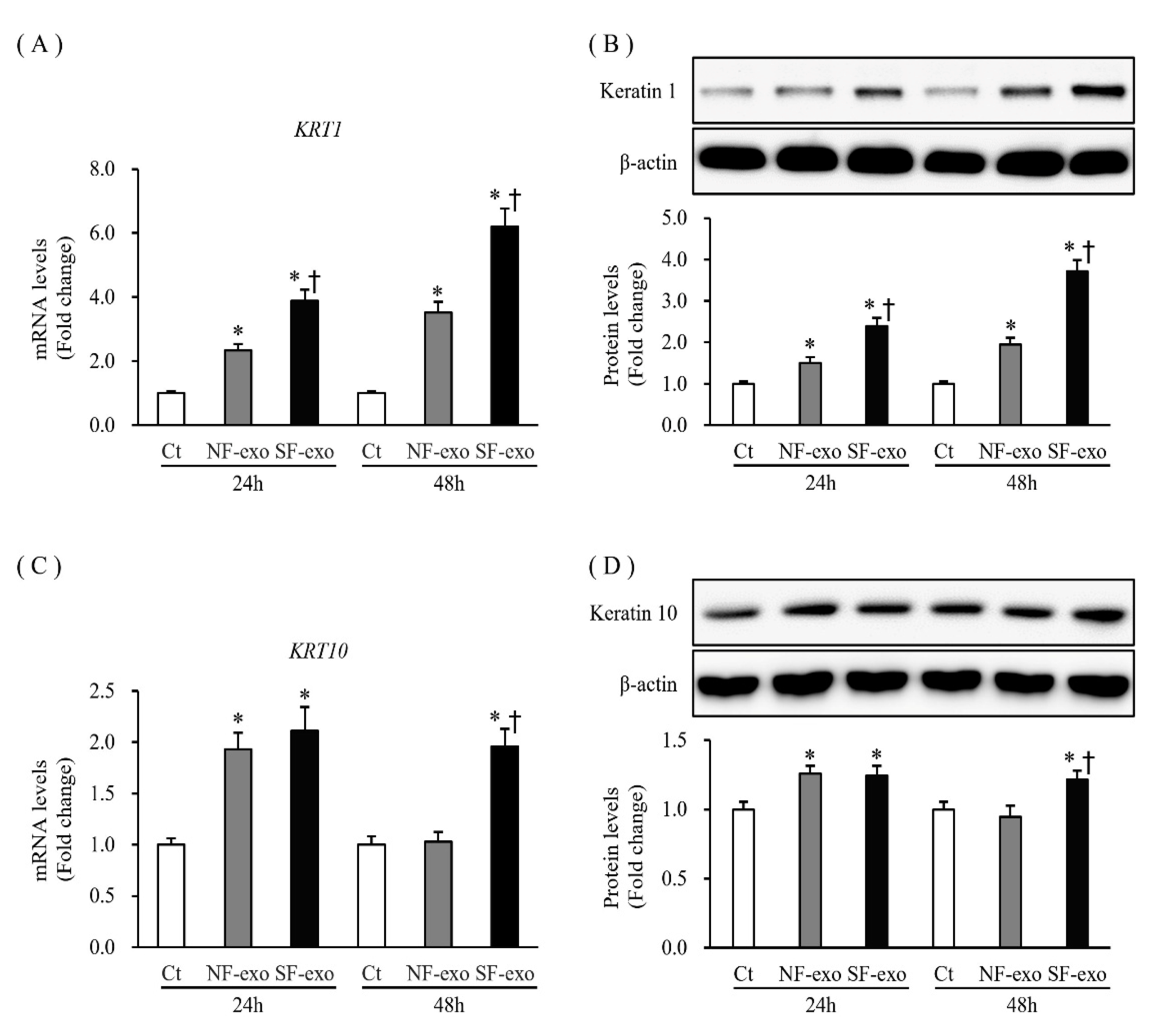
2.4. Effects of NF- and HTSF-Exosomes on the Expression of Differentiation Markers in NHKs

KRT1 and KRT10 are early differentiation markers of keratinocytes [28]. The mRNA and protein expression of KRT1 increased significantly at 24 and 48 h after treatment with NF- and HTSF-exosomes, respectively, compared with that in DPBS-treated control cells. Moreover, compared with NF-exosome treatment, mRNA and protein expression of KRT1 significantly increased at 48 h after HTSF-exosome treatment (compared with that in the DPBS-treated control cells; mRNA expression increased by 2.33- and 3.89-fold at 24 h and 3.52- and 6.21-fold at 48 h after treatment with 100 μg/mL NF- and HTSF-exosomes, respectively; protein levels increased by 1.50- and 2.39-fold at 24 h and 1.94- and 3.71-fold at 48 h in response to 100 μg/mL NF- and HTSF-exosomes, respectively; p < 0.05) (Figure 4A,B; Figure S4). The mRNA and protein expression of KRT10 increased at 24 h and was unchanged at 48 h after treatment with NF-exosomes, compared with that in the DPBS-treated cells, but significantly increased at 24 h and 48 h after treatment with HTSF-exosomes, compared with that in the DPBS-treated cells (mRNA expression was 1.93- and 2.11-fold at 24 h and 1.03- and 1.96-fold at 48 h; protein levels were 1.25- and 1.24-fold at 24 h and 0.94- and 1.21-fold at 48 h in response to 100 μg/mL NF- and HTSF-exosomes, respectively; p < 0.05) (Figure 4C,D; Figure S4). These results suggest that HTSF-exosomes substantially induced the differentiation of NHKs more than NF-exosomes.

2.5. Effects of NF- and HTSF-Exosomes on the Expression of Apoptosis-Related Factors Related to Proliferation and Differentiation in NHKs

Keratinocyte apoptosis is closely involved in epidermal development; the expression of apoptotic factors also reflects the state of cellular proliferation and differentiation, including Bax, Bcl2, apoptosis signal-regulating kinase 1 (ASK1), and Notch 1 [28]. The mRNA expression and protein levels of the pro-apoptotic factor, Bax (BAX), increased significantly at 24 and 48 h after NF- and HTSF-exosome treatment, respectively (mRNA levels increased by 1.79- and 1.85-fold at 24 h and 1.96- and 1.85-fold at 48 h after treatment with 100 μg/mL NF- and HTSF-exosomes, respectively; protein levels increased by 1.2- and 1.34-fold at 24 h and 1.41- and 1.28-fold at 48 h after treatment with 100 μg/mL NF- and HTSF-exosomes, respectively; p < 0.05) (Figure 5A,B; Figure S5). The expression of the anti-apoptotic factor, Bcl2, at the mRNA and protein levels increased significantly at 24 and 48 h (mRNA expression increased by 1.95- and 2.27-fold at 24 h and 2.13- and 2.03-fold at 48 h after treatment with 100 μg/mL NF- and HTSF-exosomes, respectively; protein levels increased by 1.53- and 1.86-fold at 24 h and 1.41- and 1.28-fold at 48 h after treatment with 100 μg/mL NF- and HTSF-exosomes, respectively; p < 0.05) (Figure 5C,D; Figure S5). The mRNA and protein expression of ASK1 (MAP3K5) increased significantly at 24 h (mRNA levels increased by 3.2- and 2.93-fold after treatment with 100 μg/mL NF- and HTSF-exosomes, respectively; protein levels increased by 2.36- and 2.12-fold after treatment with 100 μg/mL NF- and HTSF-exosomes, respectively; p < 0.05) (Figure 5E,F; Figure S5). The mRNA and protein expression of ASK1 (MAP3K5) increased at 48 h after treatment with NF-exosomes, but decreased with HTSF-exosome treatment at 48 h (mRNA levels were 2.02- and 0.36-fold and protein levels were 1.83- and 0.71-fold in response to 100 μg/mL NF- and HTSF-exosomes, respectively) (Figure 5E,F; Figure S5). The mRNA and protein expression of caspase 14 (CASP 14) decreased significantly at 24 h after treatment with NF- or HTSF-exosomes, compared with that in the control cells (mRNA expression decreased by 0.36- and 0.33-fold after treatment with 100 μg/mL NF- and HTSF-exosomes, respectively; protein expression decreased by 0.69- and 0.65-fold after treatment with 100 μg/mL NF- and HTSF-exosomes, respectively; p < 0.05) (Figure 5G,H; Figure S5). Furthermore, mRNA and protein expression of CASP 14 at 48 h increased after NF-exosome treatment, but decreased with HTSF-exosome treatment (mRNA levels were 1.96- and 0.29-fold after treatment with 100 μg/mL NF- and HTSF-exosomes, respectively; protein levels were 1.29- and 0.46-fold in response to 100 μg/mL NF- and HTSF-exosomes, respectively; p < 0.05) (Figure 5G,H; Figure S5). These results further suggest that NF- or HTSF-exosome treatment regulates the expression of apoptosis-related factors in NHKs.

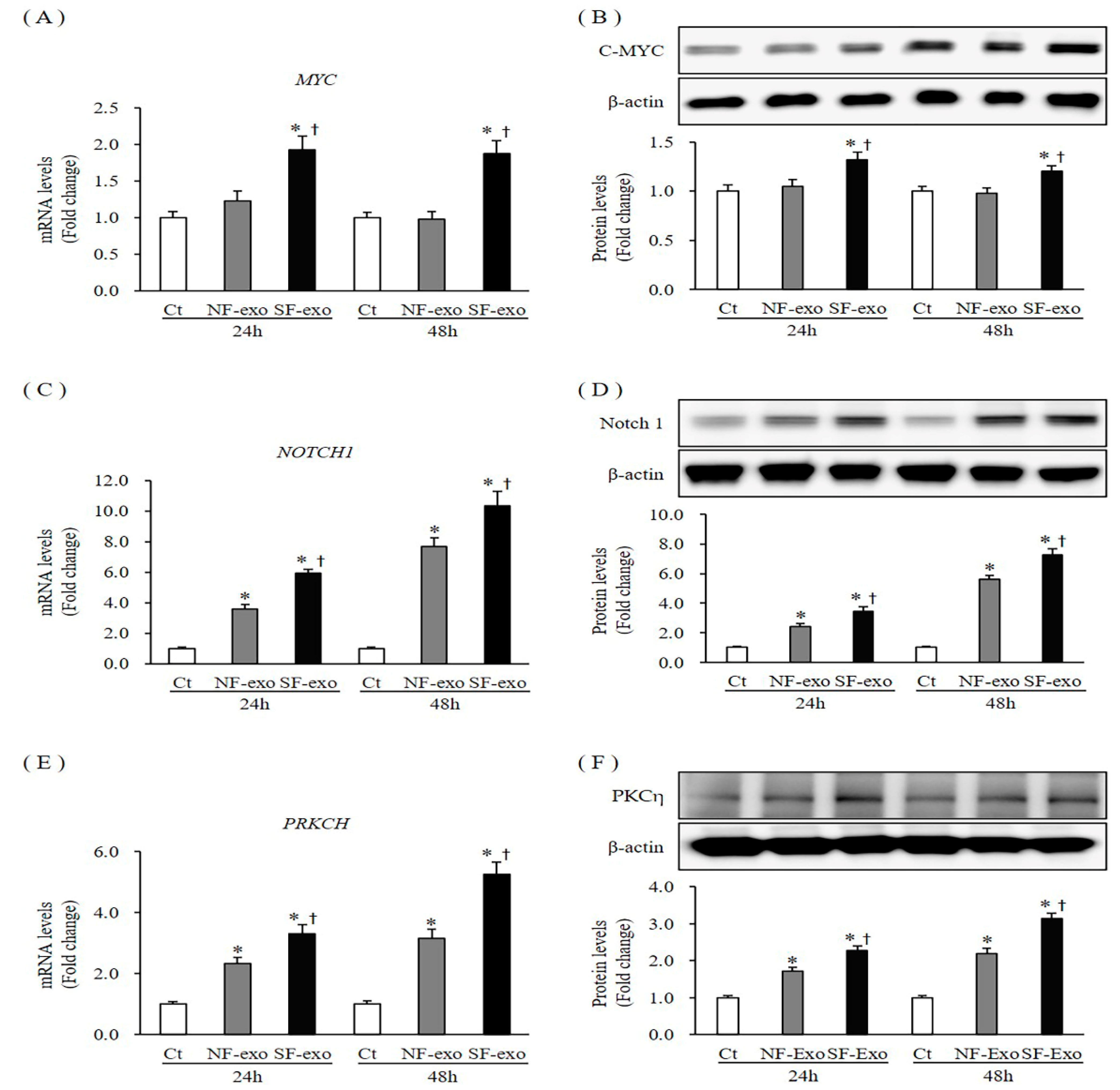
2.6. Effects of NF- and HTSF-Exosomes on the Expression of Cyclin-Dependent Kinase Inhibitors and Their Regulators in NHKs

p21 and p27 are cyclin-dependent kinase inhibitors that regulate the cell cycle and are associated with the proliferation and differentiation of keratinocyte phenotypes [29]. The p21 (CNKN1A) mRNA and protein expression levels increased significantly at 24 and 48 h (mRNA expression increased by 1.75- and 1.82-fold after 24 h and 1.97- and 2.28-fold after 48 h at 100 μg/mL, respectively; protein levels increased by 1.23- and 1.27-fold after 24 h and 1.66- and 1.67-fold after 48 h at 100 μg/mL, respectively; p < 0.05) (Figure 6A,B; Figure S6). However, p27 (CDKN1B) expression remained unchanged at 24 h and increased significantly at 48 h (at 24 h, mRNA expression was 1.09- and 1.10-fold and protein levels were 1.09- and 1.10-fold higher at 100 μg/mL, respectively, p > 0.05; at 48 h, mRNA expression was 1.98- and 3.17-fold and protein levels were 1.57- and 1.82-fold higher at 100 μg/mL, respectively; p < 0.05) (Figure 6C,D; Figure S6). These results indicate that NF-exosome treatment and, especially. HTSF-exosome treatment regulated the expression of p21 and p27 in NHKs.
The expression of p21 and p27 is tightly regulated by several factors, such as C-MYC, Notch1, and PKCη [30,31,32]. The C-MYC (MYC) mRNA and protein levels remained unchanged at 24 h and 48 h after treatment with 100 μg/mL NF-exosomes, compared with DPBS-treated control cells (mRNA expression was 1.13- and 0.98- fold at 24 and 48 h, protein levels were 1.04- and 0.97-fold and 24 and 48 h, respectively). However, MYC mRNA and protein levels significantly increased at 24 h and 48 h after treatment with 100 μg/mL HTSF-exosomes, compared with DPBS-treated control cells and NF-exosome treated cells (mRNA expression was 1.93- and 1.88- fold at 24 and 48 h and protein levels were 1.32- and 1.23-fold and 24 and 48 h, respectively, p < 0.05) (Figure 7A,B; Figure S7). Compared with NF-exosomes, mRNA and protein expression of MYC more significantly increased with HTSF-exosome treatment at 24 and 48 h (Figure 7A,B; Figure S7). The mRNA and protein level expression of Notch1 (NOTCH1) increased significantly at 24 and 48 h after treatment with 100 μg/mL NF- or HTSF-exosomes, respectively, compared with DPBS treatment (mRNA expression was 3.6- and 5.9-fold at 24 h and 7.69- and 10.32-fold at 48 h, respectively; protein levels were 2.39- and 3.45-fold at 24 h and 5.6- and 7.25-fold at 48 h, respectively; p < 0.05) (Figure 7C,D; Figure S7). Furthermore, compared with NF-exosomes, mRNA and protein level expression of NOTCH1 was more significantly increased by HTSF-exosome treatment at 24 and 48 h (p < 0.05) (Figure 7C,D; Figure S7). The mRNA and protein expression of PKCη (PRKCH) increased consistently from 24 to 48 h after treatment with 100 μg/mL NF- and HTSF-exosomes compared with control, respectively (mRNA expression was 2.33- and 3.31-fold at 24 h and 3.16- and 5.26-fold at 48 h, respectively; protein levels were 1.7- and 2.16-fold at 24 h and 2.19- and 3.12-fold at 48 h, respectively; p < 0.05) (Figure 7E,F; Figure S7). Compared with NF-exosomes, mRNA and protein expression of PRKCH more significantly increased with HTSF-exosome treatment at 24 and 48 h (Figure 7E,F; Figure S7). These results suggest that NF-exosome treatment and, especially, HTSF-exosome treatment upregulated the expression of C-MYC, Notch1, and PKCη in NHKs.

2.7. Effects of NF- and HTSF-Exosomes on the Epithelial–Mesenchymal Transition (EMT) in NHKs

E-cadherin, N-cadherin, vimentin, and cell migration are well-known EMT markers [33]. The mRNA and protein levels of E-cadherin (CDH1) decreased significantly at 24 and 48 h after 100 μg/mL NF-exosome treatment, compared with DPBS treatment, respectively (mRNA levels were 0.56- and 0.61-fold and protein levels were 0.70- and 0.75- fold at 24 and 48 h, respectively; p < 0.05) (Figure 8A,B; Figure S8). Moreover, the mRNA and protein levels of CDH1 further decreased with 100 μg/mL HTSF-exosome treatment at 24 and 48 h, respectively (mRNA levels were 0.29- and 0.33-fold and protein levels were 0.33- and 0.37- fold at 24 and 48 h, respectively; p < 0.05) (Figure 8A,B; Figure S8). The mRNA and protein levels of N-cadherin (CDH2) increased significantly at 24 and 48 h after 100 μg/mL NF-exosome treatment, compared with DPBS treatment, respectively (mRNA levels were 3.71- and 2.93-fold and protein levels were 2.32- and 1.53- fold at 24 and 48 h, respectively, p < 0.05) (Figure 8C,D; Figure S8). Moreover, the mRNA and protein levels of CDH2 further increased with 100 μg/mL HTSF-exosome treatment at 24 and 48 h, respectively (mRNA levels were 5.91- and 4.13-fold and protein levels were 3.42- and 2.83- fold at 24 and 48 h, respectively; p < 0.05) (Figure 8C,D; Figure S8). The mRNA and protein expression of vimentin (VIM) increased significantly at 24 and 48 h after 100 μg/mL NF-exosome treatment, compared with DPBS treatment, respectively (mRNA levels were 1.83- and 1.93-fold and protein levels were 1.25- and 1.31- fold at 24 and 48 h, respectively, p < 0.05) (Figure 8E,F; Figure S8). Moreover, the mRNA and protein expression of VIM further increased with 100 μg/mL HTSF-exosome treatment at 24 and 48 h, respectively (mRNA levels were 2.53- and 3.21-fold and protein levels were 1.54- and 1.91- fold at 24 and 48 h, respectively; p < 0.05) (Figure 8E,F; Figure S8). Cell migration also increased significantly at 24 and 48 h after 100 μg/mL NF-exosome treatment, compared with DPBS treatment, respectively (an increase of 170.3% and 191.6% at 24 h and 48 h, respectively; p < 0.05) (Figure 9A,B). Moreover, migration of NHKs further increased with 100 μg/mL HTSF-exosome treatment at 24 and 48 h, respectively (an increase of 338.6% and 358.7% at 24 and 48 h, respectively; p < 0.05) (Figure 9A,B). These results suggest that HTSF-exosome treatment strongly induced EMT, compared with NF-exosomes in NHKs.

3. Discussion

Post-burn HTS are conventionally attributed to the pathological development of dermis after injury. Wide-ranging studies have focused on the performance of fibroblasts but have not considered the role of epidermal keratinocytes. Nevertheless, fibroblast proliferation and dermal thickness increased upon the addition of HTS keratinocytes, compared with normal skin keratinocytes, in a tissue-engineered model [34]. The expression of IL-1α, which inhibits collagen metabolism, decreased and the expression of platelet-derived growth factor (PDGF), which upregulates dermal matrix, increased in the epidermis of HTS compared with normal scar tissues at 12 months post-surgery [35]. The epidermal expression of IL-1α and PDGF in HTS can predict whether the scar will maintain hypertrophic nature. These results suggest epidermis participation in post-burn HTS development. However, few studies have focused on the pathological characteristics of keratinocytes as the main components of epithelium. A few studies using immunohistochemistry staining showed that KRT6 and KRT16 (activation markers) were highly expressed in the interfollicular epidermis of hypertrophic scars than in the matched control sample from the same patients, while the expression of KRT1 and KRT10 (differentiation markers) was not reduced. However, the expression of another differentiation marker, filaggrin, increased [36]. In addition, focus on biological processes showed that keratinocytes in HTs expressed high levels of KRT5 (proliferation marker), KRT16 (activation marker), and filaggrin (differentiation marker) at one month after a burn. KRT16 and KRT17 (activation marker) were still upregulated at four months in HTS [37]. Moreover, both filaggrin and involucrin (differentiation marker) were at abnormal levels in HTS tissues [38]. Recently, we also reported that levels of KRT1, KRT6, KRT17, and involucrin increased in keratinocytes derived from HTS compared with their levels in matched NHKs [39]. Conventionally, however, the filaggrin expression is reduced or absent in hyperproliferative diseases, such as psoriasis and atopic dermatitis [40,41]. These results suggest that the epidermal keratinocytes in HTS display an activated phenotype and have an alternative differentiation program. Although epidermal activation has been observed, it is difficult to conclude whether it is only a consequence of HTS formation or a causal relationship with the pathogenesis.
Several studies have documented the roles of exosomes as a mechanism of cell-to-cell communication, especially as biomarkers and as a therapeutic target in fibrosis. Furthermore, exosomes derived from primary human lung fibroblasts (PHLFs) and the bronchoalveolar lavage fluid of patients with idiopathic pulmonary fibrosis increased the proliferation of PHLFs via Wnt family member 5A (WNT-5A) signaling [42]; exosomes released by cardiac myofibroblasts induced cardiac endothelial cell dysfunction by reduction of vascular endothelial growth factor (VEGF)-A, hypoxia-inducible factor-1α, CD31, and angiopoietin-1 gene expression, cell migration, and tube formation [24]; and exosomes derived from injured tubular epithelial cells can be transferred to recipient cells, leading to their proliferation and differentiation along with increasing the expression of α-SMA, F-actin, and type I collagen [23]. These studies indicate that the exosomes derived from pathological conditions differ from those in the physiological state and contribute to the pathology. In addition, exosomes derived from different subtypes of stem cells exhibit therapeutic potential and are expected to be an alternative means of developing strategies for curing various diseases [43]. The profibrotic potency of HTSF-exosomes was demonstrated by promotion of human NF proliferation and differentiation by induction of Smad-dependent and non-Smad-dependent signaling [26]. HTSF-exosomes contain large amounts of miRNA related to fibrosis, compared with those in NFs [26]. In addition, exosomes are reported to be involved in the development of organ fibrosis [44]. Therefore, the HTSF-exosomes can affect epidermal keratinocyte phenotypes. Notably, in the present study, the expression of KRT6, 16, and 17 was increased in NHKs after exposure to HTSF-exosomes. These events are also found in other hyperproliferative diseases, such as psoriasis [45].
In the dermal tissue, apoptosis controls the myofibroblast density to resemble ECM deposition. Therefore, the reduced apoptosis level is an important factor in HTS formation [4]. In epidermal cells, apoptosis maintains their thickness by balancing cell proliferation and differentiation through “an apoptotic gene program” [46]. For example, proliferating basal keratinocytes highly express Bcl-2, which is absent in suprabasal keratinocytes [47]; a gradient of Bax expression increases from lower to upper layers to promote cell death [48] and Bax expression is stronger in suprabasal keratinocytes than in basal keratinocytes [49]. In addition, the induction of ASK1 not only results in morphological changes in keratinocytes without any symptoms of the apoptotic phenotype, as evaluated by TUNEL staining, but also strongly expresses differentiation markers involucrin and transglutaminase-1 [50]. Caspase-14 is expressed only in the skin, unlike other known caspases that are expressed ubiquitously [51]. The accumulation of caspase-14 is associated with keratinocyte differentiation and stratum corneum formation [52]. Our results show that Bax expression was downregulated and Bcl-2, ASK1, and Caspase 14 expression was upregulated in NHKs after treatment with HTSF-exosomes, respectively. Moreover, the proliferation marker Ki-67 was strongly induced. These findings indicate that HTSF-exosomes can regulate proliferation and differentiation of NHKs.
p21 and p27 are important inhibitors of cyclin-dependent kinase, which is associated with keratinocyte differentiation, growth arrest, and terminal differentiation [29]. The downregulation of p21 expression by small interfering RNA transfection reversed the inhibition of the proliferation and migration of carcinoma cells [53]. p21 expression increased in the early stages of differentiated keratinocytes [54] and decreased in spontaneously terminal differentiated keratinocytes, whereas p27 expression remained unchanged [55]. However, compulsively increasing p21 expression inhibited cell growth and decreased expression of differentiation markers loricrin, involucrin, and KRT1, without affecting the cell cycle [55]. These studies suggest that p21 expression could correlate with onset but did not necessarily correlate with the late terminally differentiated phenotype of keratinocytes. Data presented in previous studies demonstrated that the differentiation of murine primary keratinocytes requires p27 induction [56]. A comparative analysis revealed an increased proliferation potential in both p21- and p27-deficient keratinocytes, but to a greater extent in p21-knockout than in p27-knockout keratinocytes. Moreover, the functions of p21 and p27 differ in the expression of differentiation markers in calcium-containing culture. Both p21- and p27-deficient keratinocytes decreased KRT1 expression, while p21-deficient cells alone specifically decreased loricrin and involucrin expression [29]. Therefore, the induction of NHK differentiation by HTSF-exosomes was partially mediated by the upregulation of both p21 and p27 expression.
Several signaling upstream molecules are involved in keratinocyte proliferation and differentiation, such as Notch1, Myc, and PKCη. The Notch signaling controls keratinocyte growth arrest and early differentiation [57]. A detailed in vivo study revealed that the deletion of the Notch1 gene led to dramatically increased epidermal cell proliferation with measurement of Ki-67 and KRT6. Moreover, in vitro, endogenous Notch activity and p21 protein expression increased in differentiating keratinocytes, along with increased extracellular calcium. p21 expression was induced by activated Notch1. Notch1 expression also increased in late terminal differentiating keratinocytes cultured from embryonic mouse skin, compared with those obtained from newborn mouse [31]. c-Myc is a positive regulator of keratinocyte proliferation [58]. Suppression of c-Myc activity inhibited cell proliferation and induced differentiation [59]. Conversely, the induction of c-Myc downregulated p21 expression, which may be one of the mechanisms through which c-Myc promoted cell proliferation [30]. In addition, several studies have established the relationship between PKCη and growth and differentiation of keratinocytes. In PKCη-null keratinocytes, p27 mRNA level was downregulated, and both growth arrest and terminal differentiation were delayed. The re-expression of PKCη or the inhibition of JNK/c-Jun signaling induced the upregulation of p27 mRNA, resulting in cell cycle arrest and terminal differentiation [32]. PKCη could activate Fyn, a Src kinase family member, which is required for normal keratinocyte differentiation. The overexpression of Fyn elevated the expression of p21 and p27, which in turn induced differentiation and growth arrest [60]. Therefore, the results from these studies support our results that HTSF-exosomes increase PKCη and Notch1 to regulate the expression of p21 and p27 and induce NHK differentiation.
EMT is a biophysical molecular process in which epithelial cells lose epithelial cell markers to acquire a mesenchymal cell phenotype, which is followed by an upregulation of mesenchymal cell markers, such as decreased expression of E-cadherin, increased expression of N-cadherin and vimentin, and increased migration [33]. Keratinocyte differentiation has a close relationship with EMT, which is also known as a differentiation program [61]. Previous in vivo studies have demonstrated that vimentin-deficient cells exhibited deprivation of EMT-like keratinocyte activation, poor keratinization, and slow re-epithelialization in wounds that are mediated by transdifferentiation and keratinocyte migration via Slug-EMT signaling in keratinocytes [61]. We found that HTSF-exosomes induce the upregulation of N-cadherin and vimentin expression and migration of NHKs, which is an example of exosomes promoting keratinocyte differentiation.

4. Materials and Methods

4.1. Primary Cell Culture

The NHKs used in this study were derived from skin biopsy, whereas HTSFs were isolated from burn-injured HTS tissues (size: 10–15 cm × 10–15 cm) obtained during surgical procedures and from normal skin that matched HTS tissue from four patients. The scars ranged from 1–2 years in age. This study was approved by the Hallym University Hangang Sacred Heart Hospital Institutional Review Board (HG 2019-016). Cell culture was performed as previously described [26,39]. Briefly, normal skin and HTS tissues were cut into small pieces, soaked in a Dispase II (1 U/mL; Gibco, Waltham, MA, USA) solution, and maintained at 4 °C overnight. Subsequently, the epidermis was separated from the dermis and digested with 0.125% trypsin (Biowest, Riverside, MO, USA) for 15 min at 37 °C. The separated dermises from normal and HTS tissues were digested with a Collagenase Type IV solution (500 U/mL; Gibco) at 37 °C for 30 min. Both solutions were inactivated with keratinocyte growth medium 2, containing supplements (PromoCell, Heidelberg, Germany), and Dulbecco’s modified Eagle’s medium (DMEM), containing 10% fetal bovine serum (FBS; Biowest) and 1% antibiotic-antimycotic with penicillin, streptomycin, and amphotericin B (Gibco). The solutions were then filtered and centrifuged at 300× g for 5 min. Pellets were obtained and resuspended in a complete medium, followed by culture at 37 °C in 5% CO2. The NHKs and HTSFs at passage two were used for all experiments.

4.2. Exosome Extraction and Treatment

Exosome extraction was performed as described previously [26]. The detailed procedure is as follows. HTSFs at 70% confluence were washed twice with DPBS (Biowest) and then with DMEM containing 10% Exo-depleted FBS (Gibco) for 48 h. Subsequently, the media from the cell culture were collected and centrifuged at 300× g for 10 min and at 16,000× g for 30 min to remove any remaining cell debris. The supernatants were subjected to centrifugation using Macrosep Advance Centrifugal Devices with a 100 kD omega membrane (Pall Corporation, Port Washington, NY, USA) at 4000× g for 1 h. The remaining Exo-containing solutions were collected; exosomes were isolated using an Exo-spinTM midi Purification Kit (Cell Guidance Systems, Cambridge, UK), according to the manufacturer’s instructions. Briefly, the solution was mixed with a 1/2 volume of exosome-spin buffer and incubated overnight at 4 °C. The mixture was then centrifuged at 16,000× g for 1 h, carefully aspirated, and the supernatant was discarded. The Exo-containing pellet was resuspended in 1 mL of DPBS, applied to the top of the column (100 μL/column), and centrifuged at 50× g for 60 s. PBS (200 µL) was applied to the column and centrifuged at 50× g for 60 s. The eluates obtained were considered to contain purified exosomes. The surface markers of exosomes were detected by determining the expression of CD9, CD63, and CD81 [2] using Western blotting. The total protein concentration of exosomes was quantified using a bicinchoninic acid (BCA) protein assay kit (Thermo Scientific, Waltham, MA, USA). The exosomes were stored at –80 °C until use. For the cell proliferation assay and the mRNA and protein expression analysis, NHKs were treated for 24 or 48 h with 100 μg/mL of HTSF-derived exosomes, whereas the control groups were treated with DPBS. For the migration assay, NHKs were treated with the same concentration for 24 or 48 h.

4.3. Cell Proliferation Assay

NHK proliferation was assessed using the CellTiter-Glo Luminescent Cell Viability Assay kit (Promega, Madison, WI, USA), as described previously [5]. NHKs were plated in 96-well cell culture plates at a density of 2.0 × 10⁴ cells and cultured for 24 h. They were then treated with exosomes derived from the HTSFs. After 24 or 48 h of cultivation, 100 µL of CellTiter-Glo reagent was added to the medium, mixed well, and incubated for 10 min at room temperature. Luminescence was recorded using a DTX 880 multimode detector (Beckman Coulter, Fullerton, CA, USA). Viability was calculated as follows: viability (%) = (sample luminescence − background luminescence)/(control sample luminescence − background luminescence) × 100.

4.4. Quantitative Reverse Transcription-Polymerase Chain Reaction (qRT-PCR)

NHKs were harvested 24 or 48 h after Exo treatment. The total RNA was extracted using the ReliaPrep RNA Miniprep System (Promega), according to the manufacturer’s instructions. The RNA concentration was estimated using a NanoDrop Spectrophotometer (BioTek, Winooski, VT, USA), and 4 µg of RNA was used to generate cDNA with the PrimeScript RT Master Mix (Perfect Real Time) (Takara, Shiga, Japan). Subsequently, 50 ng of cDNA, 0.5 µM primers (Table 1), and a 2× PCR premix (Takara) were used for qPCR on a LightCycler 96 system (Roche, Basel, Switzerland). The reaction conditions were as follows: initial denaturation at 95 °C for 30 s, 40 cycles of amplification at 95 °C for 5 s and 60 °C for 20 s, and extension at 72 °C for 30 s. The mRNA levels of the target genes were normalized to the level of β-actin using the 2−ΔΔCT method [62]. Each qPCR was performed in duplicate with cDNA from four different NHK cultures.

4.5. Western Blot Analysis

NHKs were harvested at 24 or 48 h after Exo treatment and lysed in a radioimmunoprecipitation assay buffer containing protease and phosphatase inhibitors (Sigma, St. Louis, MO, USA). The detailed methods have been described previously [63]. The protein concentration was measured using Pierce BCA Protein Assay Kit (Thermo Fisher Scientific). Lysates were mixed with 3× blue loading buffer (Cell Signaling Technology, Danvers, MA, USA) and heated for 3 min at 95 °C. Samples were subjected to gel electrophoresis, electro-transferred onto polyvinylidene difluoride (PVDF) membranes, and blocked with a 5% (w/v) bovine serum albumin in Tris-buffered saline containing 0.1% Tween-20 (PBST) for 1 h at room temperature. The PVDF membranes were incubated with the primary antibodies (Table 2). Secondary antibodies included horseradish peroxidase (HRP)-conjugated goat anti-rabbit IgG antibody (1:3000; Millipore, Billerica, MA, USA) and HRP-conjugated goat anti-mouse IgG antibody (1:3000; Millipore). Images were obtained using a chemiluminescence imaging system (WSE-6100; ATTO, Tokyo, Japan) and the optical density of the bands was measured using the Image J software (Version 1.53, NIH, Bethesda, MD, USA). Protein expression was normalized to that of β-actin; the ratio of Exo-treated cells to DPBS-treated control cells was calculated (DPBS-treated control cells = 1.0).

4.6. Cell Migration Assay

Cell migration was analyzed in a culture insert in a 35-mm µ-dish (ibidi GmbH, Planegg, Munich, Germany), according to the manufacturer’s instructions [64]. To eliminate the impact of cell proliferation during migration, mitomycin C (5 µg/mL; Sigma) was added to the cultures. Images of cell migration were obtained at 24 h (Exo treatment for 48 h) and 48 h (Exo treatment for 72 h) after migration under a light microscope (IX 70, Olympus, Tokyo, Japan); the number of cells that had migrated into the gap (cell-free wound) was analyzed using the Image J software v 1.53 (NIH, Bethesda, MD, USA). Exo-treated cells were compared with untreated NHKs, which served as a control, for which migration was set to 100%. Each analysis was performed in triplicate.

4.7. Statistical Analyses

All results are presented as the mean ± SD. The Mann-Whitney U test was used for making comparisons between the PBS-treated control group and the Exo-treated group. Statistical analyses were performed using PASW statistics 24 (SPSS Inc., Chicago, IL, USA). p-value < 0.05 was considered statistically significant.

5. Conclusions

We observed that exosomes derived from HTSFs dominantly promote the proliferation, differentiation, and activation of human normal keratinocytes, compared with exosomes derived from NFs. Therefore, our results indicate that HTSF-exosomes contribute to the epidermal development of HTS, where it is hyper-proliferated and hyperactivated, and has an alternative differentiation program.

Supplementary Materials

The following supporting information can be downloaded at: https://www.mdpi.com/article/10.3390/ijms24076132/s1.

Author Contributions

H.S.C.: design, investigation and writing—original draft; S.Y.L.: conceptualization, investigation and writing—original draft; S.Y.J.: investigation and data analysis; Y.S.C.: writing—review and editing; D.H.K.: supervision, writing—review and editing; C.H.S.: conceptualization, writing—review and editing. All authors have read and agreed to the published version of the manuscript.

Funding

This study was supported by the Soonchunhyang University Research Fund and Basic Science Research Program through the National Research Foundation of Korea (NRF) funded by the Ministry of Education (grant numbers 2020R1I1A3074338 and 2020R1I1A1A01074923).

Institutional Review Board Statement

This study was approved by the Hallym University Hangang Sacred Heart Hospital Institutional Review Board (HG 2019-016).

Informed Consent Statement

This study was conducted on human hypertrophic scars; informed consent was obtained from all participants.

Acknowledgments

We thank all the patients and their family members for their participation in the study. We thank the staff of the Burn Center at Hangang Sacred Heart Hospital.

Conflicts of Interest

The authors declare no potential conflict of interest.

References

  1. Gauglitz, G.G.; Korting, H.C.; Pavicic, T.; Ruzicka, T.; Jeschke, M.G. Hypertrophic scarring and keloids: Pathomechanisms and current and emerging treatment strategies. Mol. Med. 2011, 17, 113–125. [Google Scholar] [CrossRef]
  2. Zhu, Z.; Ding, J.; Tredget, E.E. The molecular basis of hypertrophic scars. Burns Trauma 2016, 4, 2. [Google Scholar] [CrossRef] [Green Version]
  3. Tredget, E.E.; Shankowsky, H.A.; Pannu, R.; Nedelec, B.; Iwashina, T.; Ghahary, A.; Taerum, T.V.; Scott, P.G. Transforming growth factor-beta in thermally injured patients with hypertrophic scars: Effects of interferon alpha-2b. Plast. Reconstr. Surg. 1998, 102, 1317–1328, discussion 1329–1330. [Google Scholar] [CrossRef]
  4. Armour, A.; Scott, P.G.; Tredget, E.E. Cellular and molecular pathology of HTS: Basis for treatment. Wound Repair Regen. 2007, 15 (Suppl. 1), S6–S17. [Google Scholar] [CrossRef]
  5. Cui, H.S.; Hong, A.R.; Kim, J.B.; Yu, J.H.; Cho, Y.S.; Joo, S.Y.; Seo, C.H. Extracorporeal shock wave therapy alters the expression of fibrosis-related molecules in fibroblast derived from human hypertrophic scar. Int. J. Mol. Sci. 2018, 19, 124. [Google Scholar] [CrossRef] [PubMed] [Green Version]
  6. Penn, J.W.; Grobbelaar, A.O.; Rolfe, K.J. The role of the TGF-β family in wound healing, burns and scarring: A review. Int. J. Burns Trauma 2012, 2, 18–28. [Google Scholar]
  7. Moulin, V.; Larochelle, S.; Langlois, C.; Thibault, I.; Lopez-Vallé, C.A.; Roy, M. Normal skin wound and hypertrophic scar myofibroblasts have differential responses to apoptotic inductors. J. Cell. Physiol. 2004, 198, 350–358. [Google Scholar] [CrossRef] [PubMed]
  8. Wassermann, R.J.; Polo, M.; Smith, P.; Wang, X.; Ko, F.; Robson, M.C. Differential production of apoptosis-modulating proteins in patients with hypertrophic burn scar. J. Surg. Res. 1998, 75, 74–80. [Google Scholar] [CrossRef] [PubMed]
  9. Pastar, I.; Stojadinovic, O.; Yin, N.C.; Ramirez, H.; Nusbaum, A.G.; Sawaya, A.; Patel, S.B.; Khalid, L.; Isseroff, R.R.; Tomic-Canic, M. Epithelialization in wound healing: A comprehensive review. Adv. Wound Care 2014, 3, 445–464. [Google Scholar] [CrossRef] [Green Version]
  10. Bader, R.A.; Kao, W.J. Modulation of the keratinocyte-fibroblast paracrine relationship with gelatin-based semi-interpenetrating networks containing bioactive factors for wound repair. J. Biomater. Sci. Polym. Ed. 2009, 20, 1005–1030. [Google Scholar] [CrossRef] [PubMed] [Green Version]
  11. Karvinen, S.; Pasonen-Seppänen, S.; Hyttinen, J.M.; Pienimäki, J.P.; Törrönen, K.; Jokela, T.A.; Tammi, M.I.; Tammi, R. Keratinocyte growth factor stimulates migration and hyaluronan synthesis in the epidermis by activation of keratinocyte hyaluronan synthases 2 and 3. J. Biol. Chem. 2003, 278, 49495–49504. [Google Scholar] [CrossRef] [PubMed] [Green Version]
  12. Gallucci, R.M.; Sloan, D.K.; Heck, J.M.; Murray, A.R.; O’Dell, S.J. Interleukin 6 indirectly induces keratinocyte migration. J. Investig. Dermatol. 2004, 122, 764–772. [Google Scholar] [CrossRef] [PubMed] [Green Version]
  13. Johnson, B.Z.; Stevenson, A.W.; Prêle, C.M.; Fear, M.W.; Wood, F.M. The role of IL-6 in skin fibrosis and cutaneous wound healing. Biomedicines 2020, 8, 101. [Google Scholar] [CrossRef] [PubMed]
  14. Campaner, A.B.; Ferreira, L.M.; Gragnani, A.; Bruder, J.M.; Cusick, J.L.; Morgan, J.R. Upregulation of TGF-beta1 expression may be necessary but is not sufficient for excessive scarring. J. Investig. Dermatol. 2006, 126, 1168–1176. [Google Scholar] [CrossRef] [PubMed] [Green Version]
  15. Tomasek, J.J.; Gabbiani, G.; Hinz, B.; Chaponnier, C.; Brown, R.A. Myofibroblasts and mechano-regulation of connective tissue remodelling. Nat. Rev. Mol. Cell Biol. 2002, 3, 349–363. [Google Scholar] [CrossRef] [PubMed]
  16. Zavadil, J.; Bitzer, M.; Liang, D.; Yang, Y.C.; Massimi, A.; Kneitz, S.; Piek, E.; Bottinger, E.P. Genetic programs of epithelial cell plasticity directed by transforming growth factor-beta. Proc. Natl. Acad. Sci. USA 2001, 98, 6686–6691. [Google Scholar] [CrossRef] [Green Version]
  17. Liarte, S.; Bernabé-García, Á.; Nicolás, F.J. Human skin keratinocytes on sustained TGF-β stimulation reveal partial EMT features and weaken growth arrest responses. Cells 2020, 9, 255. [Google Scholar] [CrossRef] [Green Version]
  18. Amendt, C.; Mann, A.; Schirmacher, P.; Blessing, M. Resistance of keratinocytes to TGFbeta-mediated growth restriction and apoptosis induction accelerates re-epithelialization in skin wounds. J. Cell Sci. 2002, 115, 2189–2198. [Google Scholar] [CrossRef]
  19. Ding, J.; Hori, K.; Zhang, R.; Marcoux, Y.; Honardoust, D.; Shankowsky, H.A.; Tredget, E.E. Stromal cell-derived factor 1 (SDF-1) and its receptor CXCR4 in the formation of postburn hypertrophic scar (HTS). Wound Repair Regen. 2011, 19, 568–578. [Google Scholar] [CrossRef]
  20. Quan, C.; Cho, M.K.; Shao, Y.; Mianecki, L.E.; Liao, E.; Perry, D.; Quan, T. Dermal fibroblast expression of stromal cell-derived factor-1 (SDF-1) promotes epidermal keratinocyte proliferation in normal and diseased skin. Protein Cell 2015, 6, 890–903. [Google Scholar] [CrossRef] [PubMed] [Green Version]
  21. Raposo, G.; Stoorvogel, W. Extracellular vesicles: Exosomes, microvesicles, and friends. J. Cell Biol. 2013, 200, 373–383. [Google Scholar] [CrossRef] [Green Version]
  22. Charrier, A.; Chen, R.; Chen, L.; Kemper, S.; Hattori, T.; Takigawa, M.; Brigstock, D.R. Exosomes mediate intercellular transfer of pro-fibrogenic connective tissue growth factor (CCN2) between hepatic stellate cells, the principal fibrotic cells in the liver. Surgery 2014, 156, 548–555. [Google Scholar] [CrossRef] [PubMed] [Green Version]
  23. Borges, F.T.; Melo, S.A.; Özdemir, B.C.; Kato, N.; Revuelta, I.; Miller, C.A.; Gattone, V.H.; LeBleu, V.S.; Kalluri, R. TGF-β1-containing exosomes from injured epithelial cells activate fibroblasts to initiate tissue regenerative responses and fibrosis. J. Am. Soc. Nephrol. 2013, 24, 385–392. [Google Scholar] [CrossRef] [PubMed] [Green Version]
  24. Ranjan, P.; Kumari, R.; Goswami, S.K.; Li, J.; Pal, H.; Suleiman, Z.; Cheng, Z.; Krishnamurthy, P.; Kishore, R.; Verma, S.K. Myofibroblast-derived exosome induce cardiac endothelial cell dysfunction. Front. Cardiovasc. Med. 2021, 8, 676267. [Google Scholar] [CrossRef]
  25. Zanotti, S.; Gibertini, S.; Blasevich, F.; Bragato, C.; Ruggieri, A.; Saredi, S.; Fabbri, M.; Bernasconi, P.; Maggi, L.; Mantegazza, R.; et al. Exosomes and exosomal miRNAs from muscle-derived fibroblasts promote skeletal muscle fibrosis. Matrix Biol. 2018, 74, 77–100. [Google Scholar] [CrossRef]
  26. Cui, H.S.; Kim, D.H.; Joo, S.Y.; Cho, Y.S.; Kim, J.B.; Seo, C.H. Exosomes derived from human hypertrophic scar fibroblasts induces smad and TAK1 signaling in normal dermal fibroblasts. Arch. Biochem. Biophys. 2022, 722, 109215. [Google Scholar] [CrossRef] [PubMed]
  27. Usui, M.L.; Mansbridge, J.N.; Carter, W.G.; Fujita, M.; Olerud, J.E. Keratinocyte migration, proliferation, and differentiation in chronic ulcers from patients with diabetes and normal wounds. J. Histochem. Cytochem. 2008, 56, 687–696. [Google Scholar] [CrossRef] [PubMed] [Green Version]
  28. Freedberg, I.M.; Tomic-Canic, M.; Komine, M.; Blumenberg, M. Keratins and the keratinocyte activation cycle. J. Investig. Dermatol. 2001, 116, 633–640. [Google Scholar] [CrossRef] [Green Version]
  29. Missero, C.; Di Cunto, F.; Kiyokawa, H.; Koff, A.; Dotto, G.P. The absence of p21Cip1/WAF1 alters keratinocyte growth and differentiation and promotes ras-tumor progression. Genes Dev. 1996, 10, 3065–3075. [Google Scholar] [CrossRef] [Green Version]
  30. Coller, H.A.; Grandori, C.; Tamayo, P.; Colbert, T.; Lander, E.S.; Eisenman, R.N.; Golub, T.R. Expression analysis with oligonucleotide microarrays reveals that MYC regulates genes involved in growth, cell cycle, signaling, and adhesion. Proc. Natl. Acad. Sci. USA 2000, 97, 3260–3265. [Google Scholar] [CrossRef] [Green Version]
  31. Okuyama, R.; Nguyen, B.C.; Talora, C.; Ogawa, E.; Tommasi di Vignano, A.; Lioumi, M.; Chiorino, G.; Tagami, H.; Woo, M.; Dotto, G.P. High commitment of embryonic keratinocytes to terminal differentiation through a Notch1-caspase 3 regulatory mechanism. Dev. Cell 2004, 6, 551–562. [Google Scholar] [CrossRef] [PubMed] [Green Version]
  32. Hara, T.; Miyazaki, M.; Hakuno, F.; Takahashi, S.; Chida, K. PKCη promotes a proliferation to differentiation switch in keratinocytes via upregulation of p27Kip1 mRNA through suppression of JNK/c-Jun signaling under stress conditions. Cell Death Dis. 2011, 2, e157. [Google Scholar] [CrossRef] [PubMed] [Green Version]
  33. Kalluri, R.; Weinberg, R.A. The basics of epithelial-mesenchymal transition. J. Clin. Investig. 2009, 119, 1420–1428. [Google Scholar] [CrossRef] [Green Version]
  34. Bellemare, J.; Roberge, C.J.; Bergeron, D.; Lopez-Vallé, C.A.; Roy, M.; Moulin, V.J. Epidermis promotes dermal fibrosis: Role in the pathogenesis of hypertrophic scars. J. Pathol. 2005, 206, 1–8. [Google Scholar] [CrossRef]
  35. Niessen, F.B.; Andriessen, M.P.; Schalkwijk, J.; Visser, L.; Timens, W. Keratinocyte-derived growth factors play a role in the formation of hypertrophic scars. J. Pathol. 2001, 194, 207–216. [Google Scholar] [CrossRef]
  36. Machesney, M.; Tidman, N.; Waseem, A.; Kirby, L.; Leigh, I. Activated keratinocytes in the epidermis of hypertrophic scars. Am. J. Pathol. 1998, 152, 1133–1141. [Google Scholar] [PubMed]
  37. Hakvoort, T.E.; Altun, V.; Ramrattan, R.S.; van der Kwast, T.H.; Benner, R.; van Zuijlen, P.P.; Vloemans, A.F.; Prens, E.P. Epidermal participation in post-burn hypertrophic scar development. Virchows Arch. 1999, 434, 221–226. [Google Scholar] [CrossRef] [PubMed]
  38. Limandjaja, G.C.; van den Broek, L.J.; Waaijman, T.; van Veen, H.A.; Everts, V.; Monstrey, S.; Scheper, R.J.; Niessen, F.B.; Gibbs, S. Increased epidermal thickness and abnormal epidermal differentiation in keloid scars. Br. J. Dermatol. 2017, 176, 116–126. [Google Scholar] [CrossRef] [Green Version]
  39. Cui, H.S.; Joo, S.Y.; Cho, Y.S.; Park, J.H.; Ro, Y.M.; Kim, J.B.; Seo, C.H. Effect of extracorporeal shock wave therapy on keratinocytes derived from human hypertrophic scars. Sci. Rep. 2021, 11, 17296. [Google Scholar] [CrossRef]
  40. Bernard, B.A.; Asselineau, D.; Schaffar-Deshayes, L.; Darmon, M.Y. Abnormal sequence of expression of differentiation markers in psoriatic epidermis: Inversion of two steps in the differentiation program? J. Investig. Dermatol. 1988, 90, 801–805. [Google Scholar] [CrossRef] [Green Version]
  41. Boguniewicz, M.; Leung, D.Y. Atopic dermatitis: A disease of altered skin barrier and immune dysregulation. Immunol. Rev. 2011, 242, 233–246. [Google Scholar] [CrossRef] [Green Version]
  42. Martin-Medina, A.; Lehmann, M.; Burgy, O.; Hermann, S.; Baarsma, H.A.; Wagner, D.E.; De Santis, M.M.; Ciolek, F.; Hofer, T.P.; Frankenberger, M.; et al. Increased extracellular vesicles mediate WNT5A signaling in idiopathic pulmonary fibrosis. Am. J. Respir. Crit. Care Med. 2018, 198, 1527–1538. [Google Scholar] [CrossRef] [PubMed]
  43. Han, C.; Sun, X.; Liu, L.; Jiang, H.; Shen, Y.; Xu, X.; Li, J.; Zhang, G.; Huang, J.; Lin, Z.; et al. Exosomes and their therapeutic potentials of stem cells. Stem Cells Int. 2016, 2016, 7653489. [Google Scholar] [CrossRef] [PubMed] [Green Version]
  44. Qin, X.J.; Zhang, J.X.; Wang, R.L. Exosomes as mediators and biomarkers in fibrosis. Biomark. Med. 2020, 14, 697–712. [Google Scholar] [CrossRef]
  45. Zhang, X.; Yin, M.; Zhang, L.J. Keratin 6, 16 and 17-Critical Barrier Alarmin Molecules in Skin Wounds and Psoriasis. Cells 2019, 8, 807. [Google Scholar] [CrossRef] [PubMed] [Green Version]
  46. Raj, D.; Brash, D.E.; Grossman, D. Keratinocyte apoptosis in epidermal development and disease. J. Investig. Dermatol. 2006, 126, 243–257. [Google Scholar] [CrossRef] [Green Version]
  47. Hockenbery, D.M.; Zutter, M.; Hickey, W.; Nahm, M.; Korsmeyer, S.J. BCL2 protein is topographically restricted in tissues characterized by apoptotic cell death. Proc. Natl. Acad. Sci. USA 1991, 88, 6961–6965. [Google Scholar] [CrossRef] [Green Version]
  48. Krajewski, S.; Krajewska, M.; Reed, J.C. Immunohistochemical analysis of in vivo patterns of Bak expression, a proapoptotic member of the Bcl-2 protein family. Cancer Res. 1996, 56, 2849–2855. [Google Scholar]
  49. Tomková, H.; Fujimoto, W.; Arata, J. Expression of the bcl-2 homologue bax in normal human skin, psoriasis vulgaris and non-melanoma skin cancers. Eur. J. Dermatol. 1998, 8, 256–260. [Google Scholar]
  50. Sayama, K.; Hanakawa, Y.; Shirakata, Y.; Yamasaki, K.; Sawada, Y.; Sun, L.; Yamanishi, K.; Ichijo, H.; Hashimoto, K. Apoptosis signal-regulating kinase 1 (ASK1) is an intracellular inducer of keratinocyte differentiation. J. Biol. Chem. 2001, 276, 999–1004. [Google Scholar] [CrossRef] [Green Version]
  51. Van de Craen, M.; Van Loo, G.; Pype, S.; Van Criekinge, W.; Van den Brande, I.; Molemans, F.; Fiers, W.; Declercq, W.; Vandenabeele, P. Identification of a new caspase homologue: Caspase-14. Cell Death Differ. 1998, 5, 838–846. [Google Scholar] [CrossRef] [PubMed]
  52. Eckhart, L.; Declercq, W.; Ban, J.; Rendl, M.; Lengauer, B.; Mayer, C.; Lippens, S.; Vandenabeele, P.; Tschachler, E. Terminal differentiation of human keratinocytes and stratum corneum formation is associated with caspase-14 activation. J. Investig. Dermatol. 2000, 115, 1148–1151. [Google Scholar] [CrossRef] [Green Version]
  53. Hou, P.F.; Jiang, T.; Chen, F.; Shi, P.C.; Li, H.Q.; Bai, J.; Song, J. KIF4A facilitates cell proliferation via induction of p21-mediated cell cycle progression and promotes metastasis in colorectal cancer. Cell Death Dis. 2018, 9, 477. [Google Scholar] [CrossRef] [Green Version]
  54. Missero, C.; Calautti, E.; Eckner, R.; Chin, J.; Tsai, L.H.; Livingston, D.M.; Dotto, G.P. Involvement of the cell-cycle inhibitor Cip1/WAF1 and the E1A-associated p300 protein in terminal differentiation. Proc. Natl. Acad. Sci. USA 1995, 92, 5451–5455. [Google Scholar] [CrossRef] [Green Version]
  55. Di Cunto, F.; Topley, G.; Calautti, E.; Hsiao, J.; Ong, L.; Seth, P.K.; Dotto, G.P. Inhibitory function of p21Cip1/WAF1 in differentiation of primary mouse keratinocytes independent of cell cycle control. Science 1998, 280, 1069–1072. [Google Scholar] [CrossRef] [PubMed]
  56. Hauser, P.J.; Agrawal, D.; Flanagan, M.; Pledger, W.J. The role of p27Kip1 in the in vitro differentiation of murine keratinocytes. Cell Growth Differ. 1997, 8, 203–211. [Google Scholar]
  57. Rangarajan, A.; Talora, C.; Okuyama, R.; Nicolas, M.; Mammucari, C.; Oh, H.; Aster, J.C.; Krishna, S.; Metzger, D.; Chambon, P.; et al. Notch signaling is a direct determinant of keratinocyte growth arrest and entry into differentiation. EMBO J. 2001, 20, 3427–3436. [Google Scholar] [CrossRef] [Green Version]
  58. Waikel, R.L.; Kawachi, Y.; Waikel, P.A.; Wang, X.J.; Roop, D.R. Deregulated expression of c-Myc depletes epidermal stem cells. Nat. Genet. 2001, 28, 165–168. [Google Scholar] [CrossRef]
  59. Descargues, P.; Sil, A.K.; Karin, M. IKKalpha, a critical regulator of epidermal differentiation and a suppressor of skin cancer. EMBO J. 2008, 27, 2639–2647. [Google Scholar] [CrossRef] [Green Version]
  60. Cabodi, S.; Calautti, E.; Talora, C.; Kuroki, T.; Stein, P.L.; Dotto, G.P. A PKC-eta/Fyn-dependent pathway leading to keratinocyte growth arrest and differentiation. Mol. Cell 2000, 6, 1121–1129. [Google Scholar] [CrossRef] [PubMed]
  61. Cheng, F.; Shen, Y.; Mohanasundaram, P.; Lindström, M.; Ivaska, J.; Ny, T.; Eriksson, J.E. Vimentin coordinates fibroblast proliferation and keratinocyte differentiation in wound healing via TGF-β-Slug signaling. Proc. Natl. Acad. Sci. USA 2016, 113, E4320–E4327. [Google Scholar] [CrossRef] [PubMed] [Green Version]
  62. Livak, K.J.; Schmittgen, T.D. Analysis of relative gene expression data using real-time quantitative PCR and the 2(-Delta Delta C(T)) Method. Methods 2001, 25, 402–408. [Google Scholar] [CrossRef] [PubMed]
  63. Cui, H.S.; Cho, Y.S.; Joo, S.Y.; Mun, C.H.; Seo, C.H.; Kim, J.B. Wound healing potential of low temperature plasma in human primary epidermal keratinocytes. Tissue Eng. Regen. Med. 2019, 16, 585–593. [Google Scholar] [CrossRef] [PubMed]
  64. Cui, H.S.; Joo, S.Y.; Cho, Y.S.; Kim, J.B.; Seo, C.H. CPEB1 or CPEB4 knockdown suppresses the TAK1 and Smad signalings in THP-1 macrophage-like cells and dermal fibroblasts. Arch. Biochem. Biophys. 2020, 683, 108322. [Google Scholar] [CrossRef]
Figure 1. Expression of exosome surface markers. Western blot analyses of CD 9 (A), CD 63 (B), and CD 81 (C) expression in normal fibroblast (NF)- and hypertrophic scar fibroblast (HTSF)-exosomes. Exosomes were isolated from the serum-free culture media of fibroblasts derived from post-burn HTS. Western blot analyses were performed in triplicate.
Ijms 24 06132 g001
Figure 2. Cell proliferation and expression of proliferation markers in normal human keratinocytes (NHKs) after NF- and HTSF-exosome treatment. NHK proliferation increased significantly at 24 and 48 h after treatment with 100 μg/mL NF- and HTSF-exosomes, respectively, compared with Dulbecco phosphate-buffered saline (DPBS)-treated control cells; there was a significant difference between NF- and HTSF-exosome treatment at 48 h (A). For the fold change, control cells were marked by a value of 100%. The cell proliferation assay was performed in triplicate. Protein expression of Ki-67 (B) and the mRNA and protein expression of keratin 14 (KRT14; C,D) in NHKs increased significantly at 24 and 48 h after treatment with 100 μg/mL HTSF-exosomes, except for NF-exosomes at 48 h, compared with that in the control cells. * p < 0.05 for NF- or HTSF-exosome-treated cells versus the corresponding matched control cells. p < 0.05 for HTSF-exosome-treated cells versus NF-exosome-treated cells. Data represent the mean ± standard deviation (SD); n = 4. Ct, DPBS-treated control cells; NHK, normal human keratinocytes; NF-exo, normal tissue-derived fibroblast exosomes; SF-exo, hypertrophic scar tissue-derived fibroblast exosomes.
Ijms 24 06132 g002
Figure 3. Expression of activation markers in NHKs after NF- and HTSF-exosome treatment. The mRNA and protein expression of keratin 6 (KRT6; A,B) in NHKs increased significantly at 24 h. However, these levels were unchanged at 48 h after treatment with 100 μg/mL NF-exosomes but increased in NHKs when treated with HTSF-exosomes compared with that in the control cells. The mRNA and protein expression of KRT16 (C,D) increased at 24 h and decreased at 48 h after treatment with 100 μg/mL NF-exosomes but increased when treated with HTSF-exosomes. The mRNA and protein levels of KRT17 (E,F) in NHKs decreased at 24 h after treatment with NF- and HTSF-exosomes. The mRNA and protein levels of KRT17 decreased and increased at 48 h after treatment with NF- and HTSF-exosomes, respectively, compared with those in control cells. * p < 0.05 for NF- or HTSF-exosome-treated cells versus the corresponding matched control cells. p < 0.05 for HTSF-exosome-treated cells versus NF-exosome-treated cells. Data represent the mean ± SD; n = 4. Ct, DPBS-treated control cells; NHK, normal human keratinocytes; NF, normal tissue-derived fibroblast; HTSF, hypertrophic scar tissue-derived fibroblasts.
Ijms 24 06132 g003
Figure 4. Expression of differentiation markers in NHKs after NF- and HTSF-exosome treatment. The mRNA and protein expression of KRT1 (A,B) in NHKs increased significantly at 24 and 48 h after treatment with 100 μg/mL NF- or HTSF-exosomes, compared with that in the control cells; mRNA and protein expression of KRT1 significantly increased after treatment with HTSF-exosomes compared with NF-exosomes. The mRNA and protein expression of KRT10 (C,D) in NHKs increased and was unchanged at 24 h and 48 h after treatment with 100 μg/mL NF-exosomes, respectively; however, mRNA and protein expression of KRT10 increased at 24 h and 48 h after treatment with 100 μg/mL HTSF-exosomes, compared with that in control cells. * p < 0.05 for NF- or HTSF-exosome-treated cells versus the corresponding matched control cells. p < 0.05 for HTSF-exosome-treated cells versus NF-exosome-treated cells. Data represent the mean ± SD; n = 4. Ct, DPBS-treated control cells; NHK, normal human keratinocytes; NF, normal tissue-derived fibroblasts; HTSF, hypertrophic scar tissue-derived fibroblasts.
Ijms 24 06132 g004
Figure 5. Expression of apoptosis-related factors in NHKs after NF- or HTSF-exosome treatment. The mRNA and protein expression of Bax (BAX) (A,B) and Bcl2 (C,D) in NHKs increased significantly at 24 and 48 h after treatment with 100 μg/mL NF- or HTSF-exosomes, compared with that in the control cells. The mRNA and protein expression of ASK1 (MAP3K5) (E,F) in NHKs increased significantly at 24 h after treatment with 100 μg/mL NF- or HTSF-exosomes, compared with that in the control cells. The mRNA and protein expression of MAP3K5 increased and decreased at 48 h after treatment with NF- and HTSF-exosomes, respectively (E,F). The mRNA and protein expression of caspase 14 (CASP14) (G,H) in NHKs decreased significantly at 24 h after treatment with 100 μg/mL NF- or HTSF-exosomes, respectively, compared with that in the control cells. The mRNA and protein expression of CASP14 increased and decreased at 48 h after treatment with NF- and HTSF-exosomes, respectively (G,H). * p < 0.05 for NF- or HTSF-exosome-treated cells versus the corresponding matched control cells. p < 0.05 for HTSF-exosome-treated cells versus NF-exosome-treated cells. Data represent the mean ± SD; n = 4. Ct, DPBS-treated control cells; NHK, normal human keratinocytes; NF, normal tissue-derived fibroblasts; HTSF, hypertrophic scar tissue-derived fibroblasts.
Ijms 24 06132 g005
Figure 6. Expression of cyclin-dependent kinase inhibitors in NHKs after NF- or HTSF-exosome treatment. The mRNA and protein expression of p21 (CDKN1A) (A,B) in NHKs increased significantly at 24 and 48 h after treatment with 100 μg/mL NF- or HTSF-exosome treatment but decreased with HTSF-exosome treatment at 48 h compared with that in the control cells. The mRNA and protein expression of p27 (CDKN1B) (C,D) in NHKs increased significantly at 24 h and was unchanged at 48 h after treatment with 100 μg/mL NF- or HTSF-exosomes, respectively, compared with that in the control cells. * p < 0.05 for NF- or HTSF-exosome-treated cells versus the corresponding matched control cells. p < 0.05 for HTSF-exosome-treated cells versus NF-exosome-treated cells. Data represented with mean ± SD; n = 4. Ct, DPBS-treated control cells; NHK, normal human keratinocytes; NF, normal tissue-derived fibroblasts; HTSF, hypertrophic scar tissue-derived fibroblasts.
Ijms 24 06132 g006
Figure 7. Expression of the regulators of the cyclin-dependent kinase inhibitor in NHKs after NF-and HTSF-exosome treatment. The mRNA and protein expression of C-MYC (A,B) in NHKs increased significantly at 24 and 48 h after treatment with 100 μg/mL HTSF-exosomes but remained unchanged after treatment with NF-exosomes, compared with that in the control cells. The mRNA and protein expression of Notch1 (C,D) and PKCη (E,F) increased significantly at 24 and 48 h after treatment with 100 μg/mL NF- and HTSF-exosomes, compared with that in the control cells. The mRNA and protein expression of Notch1 and PKCη increased to a greater degree with HTSF-exosome treatment than with NF-exosome treatment. * p < 0.05 for NF- or HTSF- exosome-treated cells versus the corresponding matched control cells. p < 0.05 for HTSF-exosome-treated cells versus NF-exosome-treated cells. Data represent the mean ± SD; n = 4. Ct, DPBS-treated control cells; NHK, normal human keratinocytes; NF, normal tissue-derived fibroblasts; HTSF, hypertrophic scar tissue-derived fibroblasts.
Ijms 24 06132 g007
Figure 8. Expression of EMT markers in NHKs after NF- and HTSF-exosome treatment. The mRNA and protein expression of E-cadherin (A,B) in NHKs decreased significantly at 24 and 48 h after treatment with 100 μg/mL NF-exosome, compared with that in the control cells. They further decreased with HTSF-exosome treatment compared with NF-exosome treatment at 24 and 48 h (A,B). The mRNA and protein expression of N-cadherin (C,D) and vimentin (E,F) increased significantly at 24 and 48 h after treatment with 100 μg/mL NF-exosomes, compared with that in the control cells. They were further increased with HTSF-exosome treatment compared with NF-exosomes at 24 and 48 h (CF). * p < 0.05 for NF- or HTSF-exosome-treated cells versus the corresponding matched control cells. p < 0.05 for HTSF-exosome-treated cells versus NF-exosome-treated cells. Data represent the mean ± SD; n = 4. EMT, epithelial–mesenchymal transition; Ct, DPBS-treated control cells; NHK, normal human keratinocytes; NF, normal tissue-derived fibroblasts; HTSF, hypertrophic scar tissue-derived fibroblasts.
Ijms 24 06132 g008
Figure 9. HTSF-exosome treatment dominantly increased the migration of NHKs. NHKs were treated with 100 μg/mL of NF- or HTSF-exosomes, and the control cells were treated with DPBS. After 24 and 48 h, migration of the cells was photographed (scale bar: 500 μm) (A). The quantitative analysis of cell migration was expressed as a percentage relative to the control cells (B). Ct, DPBS-treated control cells. * p < 0.05 for NF- or HTSF-exosome-treated cells versus the corresponding matched control cells. p < 0.05 for HTSF-exosome-treated cells versus NF-exosome-treated cells. Data represent the mean ± SD; n = 4. EMT, epithelial–mesenchymal transition; Ct, DPBS-treated control cells; NHK, normal human keratinocytes; NF, normal tissue-derived fibroblasts; HTSF, hypertrophic scar tissue-derived fibroblasts.
Ijms 24 06132 g009
Table 1. Sequences of primers used in real-time PCR.
GeneAccession NoForward (5′ → 3′)Reverse (5′ → 3′)
KRT1NM_006121.4TGGATGGTGCTTATATGACGACAACTCTGCTTGGTAG
KRT6ANM_005554.4TGAAGAAGGATGTGGATGATCATACAAGGCTCTCAG
KRT10NM_000421.5GATTCTCAACCTAACAACTGGCTACCTCATTCTCATACT
KRT14NM_000526GCTGAGATCAAAGACTACAAGAAGGACATTGGCATTG
KRT16NM_005557.4CCTACTTCAAGACCATCGCCTGGCATTGTCAATCTG
KRT17NM_000422.3ATCCTGCTGGATGTGAAGACGCTCCACAATGGTACGCACCTGAC
MAP3K5NM_005923.4CGTGAGCACGCTCAGTTCTATTCCGAACCAATTCTTCCAG
NOTCH1NM_017617.5AGCCTCAACGGGTACAAGTTGACACAAGGGTTGGATTC
CASP14NM_012114.3CCTGTTGTCACCTTGCTATGTCCTTGCCTCTGTCTTAC
BAXNM_001291428.2CCTTTTGCTTCAGGGTTTCACCATGTTACTGTCCAGTTCG
BCL2NM_000633.3TGCGGCCTCTGTTTGATTTAGGCATGTTGACTTCACTTGT
CDKN1ANM_078467.3TAGGCGGTTGAATGAGAGAAAGGAGAACACGGGATG
CDKN1BNM_004064.5GCAGGAATAAGGAAGCGAGGGAACCGTCTGAAACAT
PRKCHNM_006255CGCCATCTTGAGACATCCTTTTCTCGGGATTTGATTCTGG
VIMNM_003380.5AAAGCGTGGCTGCCAAGAAACCTGTCTCCGGTACTCGTTTGA
CDH1NM_004360.5GCAGACCTTCCTCCCAATACTGGGTCGTTGTACTGAATGG
CDH2NM_001792.5CCACCTTA AAATCTGCAGGCCCATGTTACTGTCCAGTTCG
MYCNM_002467.6CCTGGTGCTCCATGAGGAGACCAGACTCTGACCTTTTGCCAGG
GAPDHNM_ 002046.7CATGAGAAGTATGACAACAGCCTAGTCCTTCCACGATACCAAAGT
Table 2. Primary antibodies used in Western blot analysis.
TargetHostDilutionCompany (Cat. No.)
CD 9Mouse1:250Invitrogen (10626D), Waltham, MA USA
CD 63Rabbit1:1000System Biosciences (EXOAB-CD63A-1), Palo Alto, CA, USA
CD 81Mouse1:250Invitrogen (10630D), Waltham, MA, USA
Ki-67Rabbit1:1000Abcam (ab16667), Cambridge, UK
Keratin 1Rabbit1:1000Abcam (ab93652), Cambridge, UK
Keratin 6Mouse1:2000Abcam (ab18586), Cambridge, UK
Keratin 10Rabbit1:500Santa Cruz Technology (sc-23877), Dallas, TX, USA
Keratin 14Rabbit1:2000Abcam (ab181595), Cambridge, UK
Keratin 16Rabbit1:2000Abcam (ab76416), Cambridge, UK
Keratin 17Rabbit1:2000Abcam (ab8068), Cambridge, UK
ASK 1Rabbit1:1000Abcam (ab45178), Cambridge, UK
Notch 1Rabbit1:1000Abcam (ab52627), Cambridge, UK
Caspase 14Rabbit1:1000Abcam (ab174847), Cambridge, UK
BaxRabbit1:1000Abcam (ab199677), Cambridge, UK
Bcl2Rabbit1:1000Abcam (ab196495), Cambridge, UK
P21Rabbit1:1000Abcam (ab109199), Cambridge, UK
P27Rabbit1:1000Abcam (ab32034), Cambridge, UK
PKCηRabbit1:1000GenScript (A00906), Piscataway, NJ, USA
VimentinRabbit1:2000Abcam (ab92547), Cambridge, UK
E-cadherinRabbit1:1000Cell Signaling Technology (3195S), Danvers, MA, USA
N-cadherinMouse1:1000Thermo Fisher Scientific (33-3900), Waltham, MA, USA
MYCRabbit1:1000Cell Signaling Technology (9402), Danvers, MA, USA
β-actinRabbit1:2000Cell Signaling Technology (4967S), Danvers, MA, USA
β-actinMouse1:1000Santa Cruz Technology (sc-47778), Dallas, TX, USA
Disclaimer/Publisher’s Note: The statements, opinions and data contained in all publications are solely those of the individual author(s) and contributor(s) and not of MDPI and/or the editor(s). MDPI and/or the editor(s) disclaim responsibility for any injury to people or property resulting from any ideas, methods, instructions or products referred to in the content.

