In this article we will discuss about:- 1. Morphology of Leishmania Donovani 2. Life Cycle of Leishmania Donovani 3. Pathogenicity 4. Treatment and Prevention of Disease Caused by Leishmania Donovani.
Morphology of Leishmania Donovani:
The parasite exists in two forms:
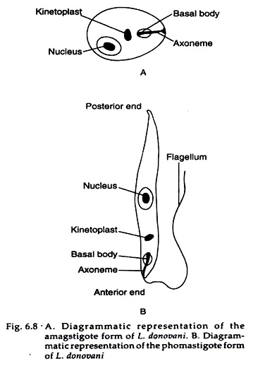
The amastigote form in man (Fig. 6.8A) and other mammals, and the promastigote form in the sand-fly.
ADVERTISEMENTS:
Amastigote form:
The amastigote form of Leishmania Donovani is also called Leishman-Donovan bodies. It is an ovoid or rounded cell, about 2.4 µm-3.5 µm wide. It is typically intracellular, being found within the cells of reticuloendothelial system (RE system) like macrophages in the spleen, monocytes, neutrophils, liver and bone marrow, and less often in other locations such as intestinal mucosa and mesenteric lymph nodes.
Smears stained with Leishman, Giemsa or Wright stains show a pale blue cytoplasm enclosed by a limiting membrane.
ADVERTISEMENTS:
The large oval or round nucleus is stained red. Lying at right angle to the nucleus is the red or purple stained kinetoplast. This kinetoplast can be seen to consist of the parabasal body and a dot-like blepharoplast, with a delicate thread connecting the two. The axoneme arising from the blepharoplast extends to the anterior tip of the cell. Alongside the kinetoplast can be seen a clear unstained vacuole.
Promastigote form:
The promastigote form of Leishmania Donovani is found in the midgut (stomach) of sand-fly. Initially it is short oval or pear-shaped forms which subsequently become long spindle-shaped cells, 15-25 µm long, carrying a single flagellum of 15-30 µm long.
Stained films show pale blue cytoplasm, with a red nucleus in the centre. The kinetoplast lies transversely near the anterior end (Fig. 6.8B). Near the root of the flagellum is present a vacuole. As the flagellum extends anteriorly without carrying back on the body, there is no undulating membrane.
Life Cycle of Leishmania Donovani:
ADVERTISEMENTS:
All leishmania pass their life cycle in two hosts. In man and some other mammals (definitive host) they occur exclusively in the amastigote form, having an ovoid body containing a nucleus and kinetoplast. In the sand-fly (intermediate host), they occur in promastigote form, with a spindle-shaped body and a single flagellum arising from the anterior end.
Definitive host:
In the body of definitive host, Leishmanias are mostly found within macrophages, monocytes, neutrophils or endothelial cells. Within these cells they multiply by binary fission, producing numerous daughter cells that distend the cells and rupture them.
The liberated daughter cells, in turn are phagocytosed by other macrophages and histiocytes, which are also killed by the parasites. By this way, the parasites eventually severely damage the reticuloendothelial (RE) system, a system that plays a critical role in the host defense.
Interestingly, amastigotes of Leishmania Donovani engulfed by neutrophils and eosinophils are killed, but in untreated cases these polymorphonuclear leucocytes have little or no effects in the eventual outcome of the disease. Due to damage of RE system, small numbers of amastigotes can be found in peripheral blood, but rarely they may be seen in faeces, urine and nasal secretions.
Intermediate host:
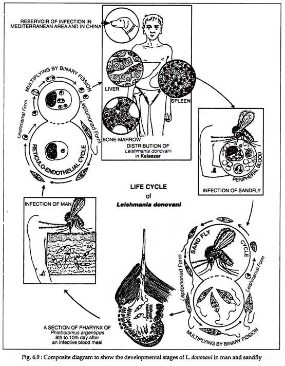
When a vector sand-fly feeds on an infected person, the amastigotes present in peripheral blood and tissue fluids enter the insect along with its blood meal. In the midgut (stomach) of the sand-fly, the amastigotes elongate and develop into the promastigote form.
The promastigotes multiply by longitudinal binary fission and reach enormous numbers. They may be seen as large rosettes, with their flagella entangled. By fourth or fifth day after feeding the parasites move forward to oesophagus and pharynx, where they accumulate and block the passage.
Such “blocked fleas” have difficulty in sucking blood when they bite a person (host) and attempt to suck blood, plugs of adherent parasites may get dislodged from the pharynx and deposited in the punctured wound. The promastigotes so deposited are phagocytosed by macrophages inside which they change into amastigotes and start multiplying.
ADVERTISEMENTS:
These, in turn, enter the midgut of sand-fly when it bites the infected person. It takes about 6-10 days for the promastigotes to reach adequate numbers so as to block the buccal cavity and pharynx of the sand-fly. This is, therefore, the duration of the extrinsic incubation period (Fig 6.9).
Pathogenicity of Leishmania Donovani:
Mode of transmission and incubation period – The infection is transmitted by the bite of the vector sand-fly Phlebotomus argentipes. The disease is not zoonotic in India, man being the only host and reservoir.
The incubation period is usually 3 to 6 months, though occasionally it may be as short as 10 days or as long as two years. Cutaneous lesion at the site of bite of the sand-fly is not seen in Indian patients but is common in patients in Sudan and Middle East.
Pathogenesis:
The disease caused by Leishmania Donovani was first characterised in India, and it is known as Kala azar (meaning black sickness), Dum Dum fever, Burdwan fever or Tropical splenomegaly. The onset of the visceral leishmaniasis is typically insidious. The clinical illness begins with fever, which may be continuous, remittent or irregular.
Splenomegaly starts early and is progressive and massive. About 10-20% patients who recovered from kala azar may develop post kala azar dermal leishmaniasis (PKDL). The dermal lesions usually develop about a year or two after recovery from the systemic disease.
Kala azar is a reticuloendotheliosis resulting from the invasion of the reticuloendothelial system by L. donovani. Several organs are affected resulting various symptoms.
(i) Splenomegaly:
The spleen is the organ most affected. It is grossly enlarged and the capsule is frequently thickened due to perisplenitis. The amastigotes multiply enormously in the fixed macrophages to produce a blockade of the RE system.
So blood-forming organs like spleen undergoes compensatory production of macrophages and other phagocytes to the detriment of red cell production. Thus, the spleen become greatly enlarged while the patient becomes generally anaemic and emaciated.
(ii) Hepatomegaly:
The liver is enlarged, the Kuffer cells and vascular endothelial cells are heavily parasitized, but hepatocytes are not affected. Liver function is therefore not seriously affected, though prothrombin production is commonly decreased. The sinusoidal capillaries are dilated and engorged. Some degree of fatty degeneration is seen.
The bone marrow is heavily infiltrated with parasitized macrophages which may crowd out the haemopoietic tissues. Peripheral lymph nodes and lymphoid tissues of the nasopharynx and intestine are hypertrophic due to infiltration with parasitized cells. The disease progresses for several months, with periods of apyrexia followed again by fever.
The skin becomes dry, rough and darkly pigmented. The hair becomes thin and brittle. Epistaxis and bleeding gums are common. Most untreated patients die in about two years due to some inter-current disease like dysentery and invasion of secondary pathogens that the body is unable to combat.
Treatment and Prevention of Disease Caused by Leishmania Donovani:
The standard treatment is the pentavalent antimonial sodium stibogluconate given I.V. (intravenous), 600 mg daily for 6 days. An alternative is pentamidine 4 mg/kg/day given I.M. (intra muscular) for 10 days. In refractory cases, splenectomy followed by chemotherapy may succeed.
Prevention:
Prophylactic measures consist of treating all cases, eradication of the vector sand-fly and personal prophylaxis by using anti-sand-fly measures.

